Envenomations Update
June 1, 2024
Related Articles
-
Echocardiographic Estimation of Left Atrial Pressure in Atrial Fibrillation Patients
-
Philadelphia Jury Awards $6.8M After Hospital Fails to Find Stomach Perforation
-
Pennsylvania Court Affirms $8 Million Verdict for Failure To Repair Uterine Artery
-
Older Physicians May Need Attention to Ensure Patient Safety
-
Documentation Huddles Improve Quality and Safety
AUTHOR
Rossi Brown, DO, ArnotHealth, Core Faculty Emergency Medicine Residency, Elmira, NY
PEER REVIEWER
Larissa I. Velez, MD, Associate Dean for Graduate Medical Education, Professor and Vice Chair for Education, Michael P. Wainscott Professorship in Emergency Medicine, Department of Emergency Medicine, UT Southwestern Medical Center, Dallas
EXECUTIVE SUMMARY
- Brown recluse spider bites can cause local erythema and sometimes develop into necrotic wounds, and infrequently develop systemic complications, including hemolysis.
- Black widow spider bites can have local erythema and diaphoresis and can progress to diffuse severe muscle cramping and spasms and systemic complications.
- Pit viper bites can cause local pain and ecchymosis, and they have potential for significant swelling and systemic complications, including coagulopathy and thrombocytopenia.
- Coral snake bites can cause neurologic dysfunction, including bulbar and generalized weakness and respiratory failure. The onset can sometimes be delayed.
- Arizona bark scorpion stings can cause local pain and paresthesias, skeletal neuromuscular dysfunction, cranial nerve dysfunction, and autonomic dysfunction, especially in children.
- Gila monster bites can cause local tissue damage and fractures, and sometimes can cause life-threatening systemic toxicity. The onset can sometimes be delayed.
- Hymenoptera stings can cause local reactions, large local reactions, and anaphylaxis. Massive envenomations, such as from Africanized honeybees, can lead to systemic toxicity, including organ failure, disseminated intravascular coagulation, and death.
- Caterpillar spines and hairs can cause local skin and eye reactions. Puss caterpillars can cause systemic toxicity.
- Blister beetles can cause local skin blisters and bullae and conjunctivitis. Ingestion can cause gastrointestinal bleeding and sometimes lead to renal failure.
Introduction
Envenomations can be caused by many different species, both marine and non-marine. The presentation can range from minor skin irritation to anaphylaxis, systemic illness, organ failure, and even death. Knowing which species are endemic to the area, and what the presentations of medically important envenomations will look like, can aid in recognition and timely treatment, especially when the bite or sting was unwitnessed. Treatment includes wound management, pain control, supportive care, and sometimes antivenom for systemic toxicity. Poison control centers and medical toxicologists are great resources that can help in treatment and disposition of various envenomations, including ones from exotic species.
This article will give an overview of medically important non-marine envenomations in the United States, including their clinical manifestations, treatment, and disposition.
Spiders
Of the thousands of spider species, most are not able to penetrate human skin and pose little or no effect on human tissues.1 Additionally, most spiders only bite humans in extreme circumstances. In the United States, two species of spiders can result in clinically significant envenomation: brown recluse and black widow spiders. (See Table 1.)
Table 1. Recluse vs. Black Widow Spider Bites |
||
Species |
Bite/Envenomation |
Treatment |
Brown recluse (Loxosceles) |
|
|
Black widow (Latrodectus) |
|
|
Recluse Spiders (Loxosceles spp.)
Loxosceles spiders, also known as recluse spiders or fiddleback spiders, are known for their bites that sometimes turn necrotic. Brown recluse spiders (L. reclusa) are the most common recluse spiders in the United States.
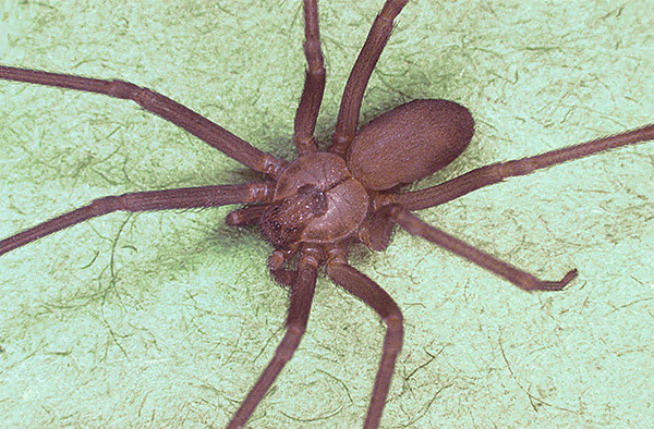
Loxosceles spiders often are identified by the violin or fiddle appearance on their back. (See Figure 1.) However, the violin marking may be absent in juvenile spiders and in some species of Loxosceles.2 Additionally, some non-venomous spiders have markings that can look like a violin and can be misidentified as a recluse spider. Other brown spiders, regardless of markings, can be mistaken for brown recluse spiders.3
Figure 1. Brown Recluse Spider |
 |
Source: CDC/Margaret Parsons |
The best way to identify Loxosceles spiders is to count the number of eyes. Most spiders have eight eyes, but the Loxosceles have six eyes. Unfortunately, most spiders are not captured for identification following a bite, and most individuals do not recall the number of eyes when describing the appearance, making confirmation of the type of spider difficult.
Endemic areas in the United States include the central/midwestern states and the southern states, excluding Florida. They usually are found in dark, quiet, dry areas of homes, such as attics, basements, and closets. They also are found outdoors in places such as wood piles.4
Clinical Manifestations
The venom of Loxosceles spiders contains cytotoxic enzymes, including phospholipase-D and sphingomyelinase.5 Bites usually are painless. Mild erythema can develop within the first eight hours, and then it fades over days to weeks. Sometimes, wounds will turn necrotic. It is estimated that 10% of bites will become necrotic, but this estimate likely is falsely elevated because of necrotic lesions being incorrectly attributed to Loxosceles spider bites.6
Wounds that turn necrotic start with pain and erythema within hours at the bite site and progress to a hemorrhagic blister surrounded by blanched skin and outer erythema. By day 3-4, the hemorrhagic blister will become ecchymotic. This gives a “red, white, and blue” appearance (outer erythema, inner blanching, and central ecchymosis). The central area may become necrotic and develop an eschar by day 5-7. The wound then will ulcerate and heal slowly. Some wounds may require skin grafting.5
Systemic symptoms are rare with United States Loxosceles spider bites. Hemolysis can occur one to three days (up to seven days) after a bite. Hemolysis is more common in children. Fever, chills, vomiting, and arthralgias also may occur. Thrombocytopenia, rhabdomyolysis, and renal failure have been described. Disseminated intravascular coagulation (DIC) and death are rare.4,5
Diagnosis is suspected based on history and physical examination. No laboratory tests are readily available for detecting spider venom. A definitive diagnosis requires observation of the spider biting the patient, the spider being captured to confirm its identity, and a physical examination that fits with spider bite and envenomation.
When a definitive diagnosis cannot be made, consider the differential diagnosis. The patient may have been bitten or stung by any number of other insects. Many insect species are more likely to bite/sting than a spider. A bite from another spider may be incorrectly identified by the patient as a brown recluse spider. Or perhaps the bite was not seen, but rather assumed. Infections that can be misidentified as a spider bite reaction include Staphylococcus and Streptococcus skin infections, herpes zoster and herpes simplex, and an erythema migrans lesion in early Lyme disease. Dermatoses from poison ivy and poison oak may be attributed to a spider bite. Pyoderma gangrenosum also can be misidentified as a spider bite.7
A mnemonic that has been proposed to help differentiate brown recluse spider bites from other diagnoses is NOT RECLUSE. It stands for Numerous (brown recluse bites usually are singular); Occurrence (did the bite happen in a typical recluse habitat); Timing (bites do not usually happen in winter months); Red center (bites usually have a pale center); Elevated (bites are flat); Chronic (lesions do not usually last several weeks); Large (lesions are not usually > 10 cm); Ulcerates too early (bites usually ulcerate after seven days); Swollen (significant swelling is atypical); Exudative (not usually exudative).8
In patients with a suspected Loxosceles bite and systemic symptoms, tests for hemolysis (complete blood count [CBC], haptoglobin), rhabdomyolysis (serum lactate dehydrogenase [LDH], creatine kinase [CK]), renal function (complete metabolic panel [CMP]), and coagulopathy (prothrombin time [PT] and partial thromboplastin time [PTT]) are recommended. Further work-up may be indicated to rule out alternate pathology and to further evaluate complications that develop.
Treatment
Management typically is supportive. This involves pain management and wound care (washing the wound, elevation, and ice packs). The cytotoxic enzyme phospholipase-D is temperature-dependent, making ice packs a good strategy to prevent the progression of necrosis.9 Antibiotics are indicated when there is a secondary wound infection. Consider tetanus prophylaxis. Surgical debridement of necrotic wounds typically occurs two to three weeks after the bite, once clear margins are established.4
Loxosceles antivenom is not available in the United States. It is available in Brazil, where Loxosceles species have higher risk of necrotic wounds and systemic illness. In Brazil, the Loxosceles antivenom is a purified equine Fab’2 in liquid suspension (“Soro antiloxoscelico”) manufactured by four large public laboratories.
Systemic complications, such as hemolytic anemia and rhabdomyolysis, are treated with standard therapies. According to a report published in 2022 of two patients bitten by L. reclusa who developed systemic illness including warm autoimmune hemolytic anemia, intravenous (IV) fluids and red blood cell transfusion are used for hemodynamic support, corticosteroids are recommended as first-line treatment, and rituximab is suggested as second-line treatment.10 A total of four case reports describe the use of therapeutic plasma exchange for refractory hemolytic anemia from brown recluse spider bites with apparent benefit.11
Some therapies have been tried in the past but lack consistent clinical benefit and could cause harm. Treatments for necrotic wounds that are not currently recommended or have insufficient evidence to recommend include dapsone, empiric antibiotics, steroids, antihistamines, hyperbaric oxygen, and early surgical intervention.2
Disposition
Hospitalization is recommended for patients with systemic manifestations after a Loxosceles bite. Consultation with the local Poison Control Center is prudent. In patients with local skin reactions, discharge is acceptable, but the patient should have outpatient follow-up for serial wound exams.2,5 For patients being discharged home, give good return precautions and ensure follow-up, since hemolytic anemia may develop days after the bite.
Black Widow Spiders (Latrodectus spp.)
Black widow spiders (Latrodectus) are found worldwide, including in most parts of the United States. In the United States, medically significant envenomations are most commonly attributed to the western black widow spider (L. hesperus) and the southern black widow spider (L. mactans).
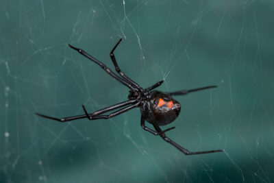
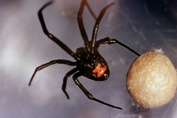
Latrodectus spiders typically are identified by the red hourglass marking on their body. (See Figure 2.) However, not all species have a perfect hourglass appearance. Sometimes, the marking appears as two separate triangles or as one triangle with a small red mark below it. Many Latrodectus spiders are black, but some are brown or red. They typically are found outdoors in garages, sheds, and woodpiles, and occasionally in gardening gloves and tools.4,12
Figure 2. Black Widow Spider |
 |
Source: CDC/Dr. Henry D. Pratt |
Clinical Manifestations
The main toxin that causes symptoms in Latrodectus spider bites is α-Latrotoxin, which causes presynaptic release of catecholamines and acetylcholine. The bite is felt as a pinprick sensation. Erythema develops within one hour of the bite. Localized diaphoresis occurs near the bite site. A target lesion with a blanched center may develop. Local paresthesias and fasciculations also may occur.
Muscle cramping and spasms frequently occur, and this pain can progress from the affected extremity to the abdomen, back, and trunk. Abdominal muscle spasms can mimic an acute abdomen. Systemic symptoms include nausea, vomiting, diaphoresis, headache, photophobia, and shortness of breath. Hypertension and tachycardia are commonly seen. Myocarditis, atrial fibrillation, pulmonary edema, and priapism are rare.
Infants and young children may present with irritability, diffuse erythema, excessive salivation, muscle tremors and weakness, tetany, and seizure-like activity.4,12 Black widow spider envenomation in pregnant women does not result in greater severity or pregnancy loss.13
There is no readily available laboratory test that can confirm envenomation. Witnessing the offending spider bite and having an examination consistent with envenomation gives the clinical diagnosis.
The differential diagnosis to consider includes an acute abdomen (severe muscle spasms can mimic an abdominal surgical emergency), acute myocardial infarction (severe muscle spasms in the chest can mimic cardiac emergency), rabies, and tetanus.12
For those with suspected Latrodectus envenomation, consider obtaining an electrocardiogram (ECG) to evaluate for dysrhythmia and a chest X-ray to evaluate for pulmonary edema. Other tests may be ordered to rule out alternate pathology, such as in the patient with suspected acute abdomen.
Treatment
Mild envenomations are limited to a local skin reaction and localized pain. Management includes oral analgesics, possibly oral muscle relaxants, and tetanus prophylaxis.14
Moderate envenomation involves spreading pain beyond the local bite site, local diaphoresis, and fasciculations. Severe envenomations involve spreading pain with spreading diaphoresis, fasciculations, muscle cramps, nausea, vomiting, and headache. Monitor airway, breathing, and circulation. IV opioids and benzodiazepines can be used for severe pain and muscle spasms.14 Caution should be used if both opioids and benzodiazepines are used on a patient, since this can lead to oversedation and respiratory depression. IV calcium gluconate had been advocated in the past, but it is no longer recommended because of its overall ineffectiveness in treating symptoms.15
Antivenom for L. mactans (Antivenin Lactrodectus mactans equine) is a treatment option for moderate to severe envenomations and is indicated when the patient is not responding to the supportive care measures mentioned earlier. However, it is in short supply, and decisions to give antivenom should be made in conjunction with a medical toxicologist.14 Antivenom has been shown to significantly reduce symptom duration in severe envenomations. Risks of antivenom include allergic reactions, anaphylaxis, and serum sickness.14,15
The antivenom dosing for adults and pediatrics is equivalent, which is 6,000 units (one vial), reconstituted with the supplied 2.5 mL sterile diluent. It can be given IV or intramuscular (IM). For IV administration, it can be further diluted with 250 mL normal saline with the infusion started at 1 mL per minute for 15 minutes and if no adverse reactions are present, the infusion rate can be increased to complete the total dose over one hour. A second and third dose can be given hourly if symptoms are persisting.14
Disposition
Patients with mild envenomations can be discharged home. Many patients with moderate to severe envenomations can be discharged home after receiving antivenom if the symptoms have resolved. The exact length of emergency department (ED) observation following antivenom administration is not established. However, one study involved an average of two hours of observation following antivenom administration.16
Hospitalization is required for those patients with ongoing or worsening systemic involvement and those who require IV pain medications.5,13 Those who receive antivenom and develop allergic symptoms should be hospitalized for ongoing observation and management. Consider admission for patients who remain hypertensive despite antivenom and pain control.15
Other Spiders
False black widow spiders (Steatoda spp.) typically produce similar but less severe symptoms than Latrodectus spiders. In the United States, S. grossa is found in coastal states. Its bites can lead to moderate to severe pain, systemic symptoms of headache and nausea, and local blistering at the bite site.17 The treatment is supportive.
Armed spiders (Phoneutria spp.) are usually found in South America. There have been reports of these spiders hiding in banana bunches during shipping and biting workers at destination sites. Bites can produce severe pain. Hypertension, tachycardia, nausea and vomiting, diaphoresis, and priapism can occur. Death from respiratory failure can occur quickly, within two to six hours, especially in children. In most cases, supportive care is sufficient. Antivenom exists, but it is unavailable in the United States.4,7
Yellow sac spiders (Cheiracanthium spp.) are found in much of the United States. They are very aggressive. Bites are painful and can lead to local erythema and edema. Nausea and headache also may occur. Treatment is supportive.4,7
Snakes
About 5,000 venomous snake bites are reported to Poison Control Centers in the United States each year. The majority are from pit vipers (Crotalinae, such as rattlesnakes and copperheads) with only 100 per year reported from coral snakes (Elapidae).18 Significant envenomation for either species can produce systemic symptoms. (See Table 2.)
Table 2. Snake Bites |
||
Species |
Envenomation |
Treatment |
Crotalinae |
|
|
Elapidae |
|
|
Pit Vipers (Crotalinae)
Members of the subfamily Crotalinae include rattlesnakes, copperheads, and water moccasins (cottonmouths). They are collectively called pit vipers. Pit vipers have heat-sensing pits behind the nostrils that help to locate and attack prey. They also have two hinged fangs that fold against the top of the mouth, unlike the elapids that have fixed fangs. Pit vipers have triangular-shaped heads, slit-like pupils, and a single row of subcaudal plates. As the name implies, rattlesnakes also have rattles.
Rattlesnakes live throughout the United States, except for Maine, Alaska, and Hawaii, and are concentrated in the southwestern United States. Copperheads are found along the eastern coast from Connecticut to Georgia and extending west to Texas. Water moccasins live along the southern United States from Virginia to Texas.
Clinical Manifestations
After a bite, pain and swelling at the bite site and ecchymosis can develop within minutes, and hemorrhagic blebs can occur within hours.
Bites to the face or neck can cause edema with resulting airway obstruction. Bites to an extremity can cause severe edema and, in rare cases, can develop into compartment syndrome. More commonly, the severe edema from envenomation is mistaken for compartment syndrome when compartment syndrome is not actually present. Increased vascular permeability and extravasation of fluid into the subcutaneous tissues can cause hypovolemia with hypotension, tachycardia, and tachypnea.
Systemic toxic effects include coagulopathy and thrombocytopenia, rhabdomyolysis, and neurotoxicity with oral paresthesias, fasciculations, and altered mental status. Other systemic symptoms include nausea, vomiting, diarrhea, chills, and generalized weakness.5,19
Bites that occur without the patient developing signs or symptoms of envenomation are called dry bites.
One in four pit viper bites is a dry bite.20 It should be noted that sometimes signs and symptoms of envenomation are delayed by several hours, so a dry bite can be diagnosed only after a period of observation.
Minimal envenomations involve only local tissue effects. Moderate envenomations include a large area of tissue effects but less than a full extremity, along with non-life-threatening systemic signs and abnormal coagulation studies without bleeding. Severe envenomations involve tissue damage to an entire extremity and tissue damage with resulting airway compromise or signs of compartment syndrome, life-threatening systemic signs, neurotoxicity, and abnormal coagulation with severe bleeding.
A history that fits with exposure to a snake, the presence of fang marks, and signs and symptoms of envenomation are needed to make the diagnosis of a snake bite with envenomation. If the first two criteria are met but without signs or symptoms of envenomation for eight to 12 hours, then a diagnosis of a dry bite can be made.
Laboratory studies should include CBC, basic metabolic panel (BMP), CK, PT and international normalized ratio (INR), fibrinogen, and D-dimer.
Treatment
Treatment should start with addressing airway, breathing, and circulation. Intubation may be required in patients with bites near the face or neck. Hypovolemia and hypotension need to be addressed early with IV fluids and vasopressors if needed. Anaphylaxis should be treated with epinephrine. Other supportive measures include pain management (avoiding nonsteroidal anti-inflammatory drugs [NSAIDs] because of coagulopathy and thrombocytopenia) and treating electrolyte derangements. A unified treatment algorithm for crotaline snakebite in the United States can be found at: https://www.ncbi.nlm.nih.gov/pmc/articles/PMC3042971/
The patient should remain calm and still, and the injured area should be kept still and elevated in both the prehospital and ED settings, using splints if available. Tourniquets to cut off arterial inflow are contraindicated since they produce damaging limb ischemia. Venous compression bandages have no proven benefit and should be avoided. Mark and time the leading edge of tenderness and swelling every 15-30 minutes. Wound management includes cleaning and covering the wound and giving tetanus prophylaxis. Wound infections are rare.
Two antivenoms to treat Crotalinae envenomations are available in the United States: Crotalidae polyvalent immune Fab (ovine) (CroFab) and Crotalidae immune F(ab’)2 (equine) (ANAVIP). A medical toxicologist consultation, such as a phone consultation with the regional Poison Control Center, is recommended before giving antivenom. See Table 3 for the CroFab treatment algorithm.21 Pregnancy is not a contraindication to antivenom administration.
Table 3. Summary of Treatment Algorithm for Crotalinae Snake Bites with CroFab Antivenom |
Step 1: Are there signs of envenomation? No
Yes
|
Step 2: Check indications for antivenom administration Is there swelling that is not minimal or is progressing, elevated PT, decreased fibrinogen or platelets, or systemic signs? No
Yes
|
Step 3: Has initial control of envenomation been obtained? Is there no progression of swelling/tenderness, labs are improving, the patient is clinically and hemodynamically stable, and neurotoxicity is improving or resolved? No
Yes
|
Step 4: Monitoring after initial control has been obtained
No
Yes
|
Adapted from: Lavonas EJ, Ruha AM, Banner W, et al. Unified treatment algorithm for the management of crotaline snakebite in the United States: Results of an evidence-informed consensus workshop. BMC Emerg Med 2011;11:2. |
A study of patients with Crotalinae bites who received CroFab found that initial control was achieved in 82.2% of patients. Neurotoxicity and thrombocytopenia were associated with difficulty in achieving initial control.22
Another study found that initial control was achieved in 87% of mild to moderate envenomations and 57% of severe envenomations. Immediate hypersensitivity reactions (anaphylactic and anaphylactoid) to antivenom occurred in 6% of patients, and delayed hypersensitivity reactions (serum sickness) developed in 5% of patients.23 Anaphylaxis can be managed using standard treatments. Serum sickness, characterized by fever, rash, and arthralgias, can be treated with antihistamines and/or NSAIDs for mild to moderate symptoms. Severe symptoms can be treated with prednisone 0.5 mg/kg to 2 mg/kg daily for seven to 10 days.
There is a risk of recurrent and late coagulopathy up to seven days after CroFab administration, as well as a risk of persistent coagulopathy despite antivenom therapy. Patients with rattlesnake or water moccasin bites should have coagulation studies rechecked at two to three days and five to seven days after the bite.24
Crotalidae immune F(ab’)2 (equine) (ANAVIP) has similar efficacy to CroFab and similar rates of adverse reactions, but with less risk of delayed coagulopathy. Ten vials of ANAVIP (for both adult and pediatric patients) can be given in moderate to severe rattlesnake envenomations, with repeat dosing of 10 vials every hour until local signs of envenomation have stopped progressing, systemic symptoms are resolved, and coagulation parameters have normalized or are trending toward normal. Although there is less risk of delayed coagulopathy with ANAVIP, it still is recommended that patients have coagulation studies rechecked five to seven days after the last antivenom dose.24
Fasciotomy of the affected limb based on physical examination findings alone is contraindicated, since the local tissue damage from venom can mimic signs and symptoms of compartment syndrome. The compartment pressure should be measured and if elevated, IV mannitol 1 g/kg to 2 g/kg over 30 minutes administered, and additional antivenom should be administered simultaneously.19 If the compartment pressure remains elevated after 60 minutes, fasciotomy should be considered, although there is no solid evidence supporting its use.19
Crotalinae venom will inactivate transfusions of clotting factors and platelets. Therefore, if coagulopathy or thrombocytopenia is present, antivenom should be administered first, and platelet/fresh frozen plasma transfusions should be reserved for severe bleeding despite antivenom.24
Disposition
Hospitalization for observation and treatment of Crotalinae bites is recommended. (See Table 3.) Follow-up to recheck coagulation studies should be arranged when it is indicated. If there was coagulopathy during the hospital stay or if the patient was bitten by a rattlesnake, instruct the patient to avoid contact sports, dental work, and surgery for two weeks. Instruct patients to watch for signs and symptoms of serum sickness if antivenom was administered.
Coral Snakes (Elapidae)
Elapids include coral snakes, mambas, and cobras. Coral snakes of the United States include the Eastern coral snakes (M. fulvius), Arizona (Sonoran) coral snakes (M. euryxanthus), and Texas coral snakes (M. tener). Eastern coral snakes are found mostly in Florida and the southern portions of its bordering states. Arizona and Texas coral snakes are found in Arizona and Texas, respectively. Mambas live in Africa, and cobras live in Africa and Southeast Asia. Mambas, cobras, and other exotic elapids can be found in the United States in zoos and occasionally as exotic pets. Coral snake bites are the most common elapid bites in the United States and will be the focus of this section.
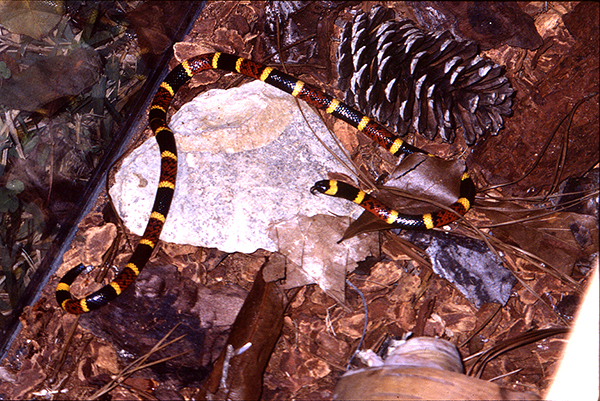
Coral snakes do not have hinged fangs but rather latch and “chew” their victims, expelling venom as they do this. Coral snakes in the United States have brightly colored rings on their skin, with red and yellow rings next to each other. (See Figure 3.) Red and black rings next to each other usually indicate a nonvenomous snake (king snake), although this rule does not hold true outside the United States. The phrase “red on yellow, kill a fellow; red on black, venom lack” is an easy way to remember venomous coral snakes vs. nonvenomous king snake species.
Figure 3. Coral Snake |
 |
Source: CDC/ Edward J. Wozniak DVM, PhD. |
Clinical Manifestations
Contrary to crotalid bites, with elapids there is minimal or no pain at the bite site, mild or no swelling, and no significant tissue damage. Fang or bite marks are not always identified. Many of these bites do not result in envenomation. When envenomation does occur, nausea, vomiting, and abdominal pain can be seen.
Coral snake venom inhibits the acetylcholine receptors at the neuromuscular junction, resulting in paresthesias, bulbar paralysis leading to double vision, ptosis, difficulty speaking, and difficulty swallowing; respiratory failure from descending muscle weakness; and generalized weakness. These neurologic effects can be delayed by up to 12 hours. Coagulopathy is not common.25
Coral snakes usually are identified when they bite a patient, since many bites occur from direct handling. Coral snakes often have to be forcibly removed since they latch on and chew their victims. When the bite is not observed, consider alternative diagnoses, such as botulism, Guillain-Barré syndrome, and myasthenia gravis.
When a snake bite is involved but there is severe local tissue damage or evidence of coagulopathy, both suggesting a non-elapid bite, consider laboratory testing as discussed in the Crotalinae section.
Pulmonary function testing, such as maximal inspiratory pressure (MIP), can be used to assess respiratory muscle weakness. Serial exams and close observation with monitoring are needed.
Treatment
Immobilize the affected limb in a position below the level of the heart and consider pressure immobilization to delay systemic absorption of venom.25 Unlike Crotalinae bites, elapid bites do not cause significant local tissue damage, and preventing spread to prevent systemic toxicity is more important. Clean the wound and provide tetanus prophylaxis.
Monitor airway, breathing, and circulation. Serial pulmonary function tests, such as the MIP (also known as the negative inspiratory force [NIF]), forced vital capacity (FVC), or maximal expiratory pressure (MEP), can guide the need for respiratory support. For example, if the MIP falls below -30 cm H2O, respiratory muscle weakness is worsening and mechanical ventilation is indicated. FVC less than 50% of predicted capacity and MEP less than 40 cm H2O also are indications for mechanical ventilation. End-tidal CO2 can detect hypoventilation, and arterial blood gases can look for impending respiratory failure.
There is a suggestion that if neurotoxicity is developing and there will be a delay in antivenom administration, one can consider anticholinesterase administration, such as neostigmine, pretreated with atropine. However, this consideration applies to coral snakes of Central and South America and does not apply to United States coral snake species.25
North American coral snake antivenom (Antivenin [Micrurus fulvius]) is indicated for all symptomatic (beyond minor pain and swelling at bite site) coral snake envenomations, except for Arizona coral snake bites (for which the antivenom does not have an effect, the envenomations are not severe, and no deaths have been reported). Three to five vials of the antivenom should be given for both adults and children.26 Adverse reactions to the antivenom, such as allergic reactions and serum sickness, are common (18%), but life-threatening reactions like angioedema and hypotension are not common.27 Allergic reactions and serum sickness can be treated with standard treatments described earlier.
Disposition
Admit all patients with coral snake bites. Asymptomatic patients may develop delayed neurologic symptoms up to 12 hours later, and, therefore, should be observed and closely monitored for 12-24 hours. Patients requiring antivenom still can have progression of neurologic symptoms and, therefore, also require admission.26
Scorpions
Scorpions are invertebrate arthropods that can be found worldwide, including the southern parts of the United States. The Arizona bark scorpion (Centruroides sculpturatus) found in Arizona, New Mexico, and parts of California and Texas, has a venom that can cause systemic signs and symptoms, and even death, especially in children. Exotic scorpions can be found in zoos and as pets.
Clinical Manifestations
A scorpion sting will cause sudden, intensive local pain followed by a tingling sensation or paresthesias. Bark scorpions do not cause local tissue destruction. Neuromuscular toxicity can develop and can be life-threatening.
Envenomation is categorized by severity. Grade I envenomation involves local pain and/or paresthesias. Tapping the sting site as the patient is looking away will increase the pain significantly. This is known as the “tap test” and is characteristic of bark scorpion stings. Grade II envenomation includes local as well as proximally spreading or remote pain and/or paresthesias. Grade III envenomation causes either cranial nerve dysfunction or skeletal neuromuscular dysfunction, with or without autonomic dysfunction. Grade IV envenomation involves both cranial nerve and skeletal neuromuscular dysfunction and also can have autonomic dysfunction.28
Cranial nerve dysfunction includes slurred speech, dysphagia, abnormal eye movements (usually slow, conjugate, wandering movements, but sometimes the classic finding of opsoclonus with rapid multidirectional movements) and blurred vision, and tongue fasciculations. Increased oral secretions and bulbar neuromuscular dysfunction can cause airway problems.
Skeletal neuromuscular dysfunction includes fasciculations, shaking and jerking of the extremities, restlessness, and alternating arching (opisthotonos) and flexion of the back. Autonomic dysfunction includes vomiting, salivation, diaphoresis, bronchoconstriction, and tachycardia.
For grade III and IV envenomations, laboratory testing for renal and hepatic function (CK), rhabdomyolysis (CK, urine myoglobin), and cardiac injury (troponin) is indicated.
The differential diagnosis, especially when the scorpion sting was unwitnessed, includes black widow spider bite, tetanus, strychnine poisoning, botulism, neuroblastoma, organophosphate toxicity, seizure, and encephalitis. Black widows can cause similar autonomic dysfunction, but without the abnormal eye movements and fasciculations. Tetanus and strychnine poisoning can cause painful muscle contractions, including opisthotonos similar to scorpion envenomation, and tetanus can involve autonomic dysfunction. However, these lack the abnormal eye movements seen with scorpion envenomation.
Botulism can cause cranial nerve dysfunction along with symmetric weakness. Unlike scorpion envenomation, botulism tends to lack fever and has symmetric neurologic symptoms, with no sensory deficits or pain. Neuroblastoma can involve abnormal eye movements and jerking of the extremities, but without pain, excessive salivation, skeletal neuromuscular dysfunction, or acute onset of symptoms. Organophosphate toxicity can cause excessive salivation and fasciculations, but it also can cause paralysis and seizures, which are not seen with scorpion envenomation.
Scorpion envenomation can have alternating arching and flexion of the back similar to a seizure, but seizing patients generally do not have normal mental status. Fever and stiffness of the neck can be seen with both scorpion envenomation and meningitis, but meningitis lacks the abnormal eye movements seen in scorpion envenomations.28
Treatment
Grade I and II envenomations can be treated with pain control, including NSAIDs, opioids, and sometimes regional anesthesia. Clean the wound and administer tetanus prophylaxis. For grade III and IV envenomations, supportive care and airway management are important along with administration of antivenom. Young children and pregnant women also should receive antivenom, Centruroides (scorpion) immune F(ab’)2 (equine) (Anascorp). Dosing for both adults and children is three vials dissolved in 20 mL to 50 mL normal saline, infused IV over 30 minutes. Patients can receive subsequent one-vial doses every 30 minutes until resolution of symptoms, up to a total of five vials.28
A study evaluating critically ill children after a scorpion sting found that Anascorp produced more rapid resolution of symptoms, resolution of symptoms in all antivenom recipients by four hours, and undetectable plasma venom concentration at one hour after treatment.29 A recent study looked at 145 patients who received Anascorp, most of whom were children, and showed rapid resolution of symptoms and no hypersensitivity reactions.30
Atropine can be considered for hypersalivation from Centruroides stings. However, some scorpions outside of the United States cause an autonomic storm, and atropine use in these cases can worsen toxicity.4
Disposition
Patients with grade I and II envenomations should be observed for four hours for progression of symptoms.28 They then can be discharged home if no progression is seen. For grade III and IV envenomations, patients who receive antivenom and have symptom resolution and vital sign normalization can be discharged home. Patients with grade III and IV envenomations with persistent signs and symptoms, persistent abnormal vital signs, complications, the need for IV analgesia, or who are intubated require admission.5
Gila Monsters (Heloderma)
Gila monsters are poisonous lizards that are found in the southwestern deserts of the United States and northern Mexico. They also are found in zoos and kept as pets. They have grooved teeth with venom that contains gilatoxin. Gila monsters have a strong bite and can be difficult to remove once latched, which can result in deep wounds and fractures.
Clinical Manifestations
Gila monsters have painful bites with tissue damage and possible underlying fractures. When envenomation occurs, systemic toxicity includes diaphoresis, vomiting, diarrhea, paresthesias, generalized weakness, and severe hypertension. Life-threatening presentations can include angioedema, volume depletion with metabolic derangements, and atrioventricular cardiac conduction disorders.31
Treatment
Local wound care and tetanus prophylaxis are needed. Control pain and remove any dislodged teeth from the wound. No antivenom is available for Heloderma envenomation, so these patients should receive supportive management. Monitor airway, breathing, and circulation. Treat cardiovascular or pulmonary complications with standard therapies.
Disposition
Patients with no systemic toxicity after several hours of observation can be discharged home. Admission is recommended for those with signs and symptoms of systemic toxicity.32
Hymenoptera
This class of insects includes the Vespidae (hornets, wasps, and yellow jackets), Apidae (honeybees and bumblebees), and Formicidae (ants, including imported fire ants).4 Hymenoptera stings cause more fatalities than any other arthropod, with about 60 deaths in the United States each year.33
Vespidae and Apidae
Clinical Manifestations
The venom of Vespidae and Apidae has many components, including phospholipase A (a strong allergen), hyaluronidase (breaks down tissue to allow venom penetration), and histamine (contributes to pain and itching). Bee venom also contains melittin (cytotoxic) and apamin (neurotoxic). Hornet and wasp venom contains acetylcholine (increases stimulation of pain nerves).34 These and other venom components are responsible for the symptoms of local reactions and anaphylaxis, and for the toxic effects of massive envenomations.
The most common reaction to Vespidae and Apidae stings is a local reaction consisting of erythema, swelling, pain, and pruritus. These develop within minutes of a sting and usually resolve over a few hours. Some people develop large local reactions, with a larger area (about 10 cm in diameter) of erythema and swelling. The erythema and swelling spread over the first 48 hours and gradually resolve over the next five to 10 days.35
Secondary bacterial infection is uncommon. However, large local reactions often are confused with secondary bacterial infections. Infection should be suspected when erythema, swelling, and pain are increasing three to five days after a sting, since large local reactions start improving after the first two days. Fever also suggests infection. Lymphangitic streaks can be seen with both infection and large local reactions.35
Anaphylaxis is a serious reaction to a Hymenoptera sting. Its symptoms can range from mild to rapidly fatal. Death can happen within minutes. Flushing, urticaria, and angioedema are common skin symptoms. Shortness of breath due to upper airway edema and wheezing due to bronchoconstriction can occur. Nausea, vomiting, diarrhea, and abdominal cramping can be seen. Hypotension and cardiovascular collapse can occur with severe anaphylaxis.
The risk of anaphylaxis from future stings is a concern for many patients. In adults with a history of anaphylaxis to Hymenoptera stings and who have venom-specific immunoglobulin E (IgE), the risk of recurrent anaphylaxis with a subsequent sting is 30% to 70%.36 Children with a history of moderate to severe anaphylaxis to a sting have a 32% chance of recurrent anaphylaxis to a sting in the future.37
Local reactions, large local reactions, and anaphylaxis can occur after one or a few stings. Massive envenomation occurs after many stings. This can cause vomiting and diarrhea, lightheadedness and syncope, involuntary muscle spasms, convulsions, and fever. Rhabdomyolysis, renal failure, liver failure, and DIC can occur.4 Honeybees and bumblebees can sting only once because their sting apparatus detaches from their body after a sting. However, hornets, wasps, and yellow jackets are able to withdraw their stinger and sting multiple times, especially when thousands of insects are swarming. Renal failure has been seen after as few as 20-30 wasp stings.38
Massive envenomations also can be seen from Africanized honeybees. Africanized honeybees, or killer bees, are found in the southern United States. They are more aggressive and can swarm and attack with more than 10 times the number of bees than with a typical North American bee. More than 50 stings in an adult and more than one sting per kilogram in a child have a risk of massive envenomation. Death can occur after hundreds of stings.5
A delayed reaction of serum sickness-like illness can occur five to 14 days after a sting. Signs and symptoms include polyarthritis, lymphadenopathy, urticaria, fever, and malaise.9 Other uncommon reactions include myocarditis, vasculitis, neuritis, and encephalitis. Although rare, these reactions can occur days to weeks after a sting.35
Laboratory studies are not usually indicated for most Hymenoptera stings. In massive envenomations, consider CMP, CK, DIC panel, troponin, and ECG testing.
Treatment
Monitor airway, breathing, and circulation. Evaluate for signs of anaphylaxis. Anaphylaxis from a sting is treated the same as other causes of anaphylaxis, including epinephrine, corticosteroids, and H1 and H2 antihistamines. (See Table 4.)
Table 4. Anaphylaxis Medications and Dosing |
||
Medication |
Adult Dosing |
Pediatric Dosing |
Epinephrine |
0.3 mg to 0.5 mg IM (1 mg/mL preparation) |
0.01 mg/kg (max 0.5 mg) |
Corticosteroid Methylprednisolone |
125 mg IV |
1 mg/kg (max 125 mg) |
H1 Antihistamine Diphenhydramine |
25 mg to 50 mg IV |
1 mg/kg (max 50 mg) |
H2 antihistamine Ranitidine |
50 mg IV |
1 mg/kg (max 50 mg) |
IM = intramuscular; IV = intravenous |
||
Bee stingers that are lodged in the skin should be flicked off immediately, or as soon after a sting as possible. Venom generally is released from the stinger within seconds of a sting, and stingers should be removed as soon as possible to prevent any remaining venom from being injected.35 Scraping off the stinger using the edge of a credit card is a safe, quick method to dislodge a stinger.
Cold compresses can be used for local reactions. Large local reactions can be treated with cold compresses, NSAIDs for pain, antihistamines and topical corticosteroids for itching, and oral prednisone.35 When secondary bacterial infection is suspected, treatment with an oral antibiotic is indicated.
Venom immunotherapy can reduce the risk of a recurrence of anaphylaxis to a sting to about 5% in both children and adults.37,39 Refer patients who develop anaphylaxis after a Hymenoptera sting to an allergist for consideration of immunotherapy.
Although bee antivenom is not yet available in the United States, Africanized honeybee antivenom was developed recently in Brazil. It currently is in a Phase II clinical trial.40
Disposition
Admission for observation should be considered for patients with massive envenomation (> 50 stings in adults and > 1 sting/kg in children). Admission criteria for anaphylaxis after a sting are the same as for other causes of anaphylaxis, such as severe initial reaction, need for multiple doses of epinephrine, history of severe asthma, or history of a biphasic reaction. Patients with other significant complications, such as myocarditis and encephalitis, also should be admitted.
Patients with uncomplicated stings can be discharged home. Prescribe an epinephrine autoinjector to those with anaphylactic reactions to a sting and refer to an allergist for follow-up.
Formicidae (Solenopsis spp.)
Imported fire ants are aggressive and will swarm and sting with little provocation. They hold onto the skin with their strong mandibles and can sting repeatedly if not removed promptly. Red imported fire ants can be found in the southern United States. Black imported fire ants are found in parts of Alabama, Mississippi, and Tennessee.41
Clinical Manifestations
Fire ant venom contains phospholipase A, hyaluronidase, and histamine, similar to bees, wasps, and hornets. The venom also contains piperidine alkaloids, which contribute to the pain of fire ant stings.34
The first symptoms to present after a sting from an imported fire ant are erythema, painful burning, and pruritus. Within 24 hours, a sterile pustule will develop for each sting. Secondary bacterial infection can occur when the pustules are opened. Sometimes they are opened when the patient scratches the area, and sometimes they are opened because the patient thinks they are infected already and need to be drained.35
Large local reactions are possible and an extreme local reaction can cause swelling severe enough to cause vascular compromise. Anaphylaxis is reported in 0.6% to 16% of patients.41 Rhabdomyolysis, acute renal failure, and cardiopulmonary symptoms resulting in death can occur with massive attacks, yet other people recover without complications.4,41 In massive attacks, consider BMP, troponin, CK, and ECG tests.
Treatment
Antihistamines and moderate-potency topical corticosteroids can provide itch relief. Oral antibiotics are indicated if a secondary bacterial infection develops. A high-potency topical steroid or oral prednisone can help with large local reactions. Anaphylaxis is treated with standard therapies. (See Table 4.)
Disposition
Patients with uncomplicated stings can be discharged home. Victims of massive attacks or those with cardiovascular or renal injury require admission.
Caterpillars
Clinical Manifestations
Caterpillars have protective hairs and spines, and the spines are attached to a venom gland. Pruritus, a rash known as “caterpillar dermatitis,” and sometimes urticaria can occur from contact with caterpillar hairs and venom.
Caterpillar hairs also can affect the eyes. Conjunctivitis, keratitis, conjunctival and iris nodules, and chorioretinopathy can develop. Ophthalmia nodosa is the term given to ocular conditions resulting from contact with caterpillar hairs.42
The puss caterpillar (Megalopyge opercularis) can cause a severe burning sensation, grid-like hemorrhagic papular rash, lymphadenopathy, and edema. Fever, hypotension, and convulsions also can occur.4
Treatment
Adhesive tape can be used to remove caterpillar hairs and spines from the skin. Cool compresses, antihistamines, and moderate-potency topical corticosteroids can be given for itching. Treatment for ophthalmia nodosa includes irrigation of the eye, topical antibiotics and steroids, sometimes systemic steroids, and removal of nodules and caterpillar hairs by an ophthalmologist.42 Pain management and supportive care are treatments for puss caterpillar reactions.
Disposition
Patients generally can be discharged home. Systemic complications, such as those seen with the puss caterpillar, may require admission.
Blister Beetles (Epicauta spp.)
Blister beetles are members of the plant-eating insect family Meloidae. Blister beetles have narrow bodies three-quarters of an inch to one and one-quarter inch in length, broad heads, and long antennae. There are four species of blister beetles common in the eastern and central United States. The bodies of living and dead blister beetles may contaminate cured hay and poison livestock.
Clinical Manifestations
Blister beetles contain cantharidin toxin. Cantharidin-containing products can be used for wart removal and have been used as an aphrodisiac. Contact with this toxin in high concentrations can produce blisters and bullae. If contact with the eye occurs, the toxin can cause severe conjunctivitis known as Nairobi eye.
Ingestion of blister beetles or cantharidin-containing products can cause vomiting, diarrhea, gastrointestinal bleeding, and abdominal pain, followed by hematuria and dysuria, oliguria, and renal failure.4 The lethal dose of cantharidin is estimated to be between 0.5 mg/kg and 1.0 mg/kg of body weight.
Treatment
Blister beetles should be removed from the body by blowing or flicking, since the toxin is released from the joints of the beetle when it is disturbed or crushed. Irrigation of the skin can remove residual toxin. Ophthalmologists can help with ocular exposures. Ingestions are treated with supportive measures.
Disposition
Admission is warranted for patients who are symptomatic after ingestion.4 Patients without systemic toxicity or symptomatic ingestion can be discharged home.
Conclusion
Envenomations can range from minor to severe and sometimes can be fatal. Practitioners should be aware of envenomation presentations, such as brown recluse vs. black widow spider bites, and crotalid vs. elapid snake bites. Practitioners should know the treatments for common or dangerous envenomations, which can range from supportive care to the use of antivenom. Contacting a medical toxicologist or regional Poison Control Center can be helpful with treatment, disposition, and knowing what to watch for. Practitioners should be aware of what venomous species live in the area where they practice. Periodic review of envenomation presentations and treatments can be helpful.
REFERENCES
- Swanson DL, Vetter RS. Bites of brown recluse spiders and suspected necrotic arachnidism. N Engl J Med 2005;352:700-707.
- Vetter RS, Swanson DL. Bites of recluse spiders. UpToDate. Updated April 5, 2024. https://www.uptodate.com/contents/bites-of-recluse-spiders
- Vetter R. Identifying and misidentifying the brown recluse spider. Dermatol Online J 1999;5:7.
- Schneir AB, Clark RF. Bites and stings. In: Tintinalli JE, Stapczynski JS, Ma OJ, et al, eds. Tintinalli’s Emergency Medicine: A Comprehensive Study Guide. 7th ed. McGraw-Hill Medical;2011:1344-1354.
- Envenomations — non-marine. In: Katz KD, O’Connor AD, eds. EMRA and ACMT Medical Toxicology Guide. Emergency Medicine Residents’ Association;2018:57-72.
- Tutrone WD, Green KM, Norris T, et al. Brown recluse spider envenomation: Dermatologic application of hyperbaric oxygen therapy. J Drugs Dermatol 2005;4:424-428.
- Vetter RS, Swanson DL. Diagnostic approach to the patient with a suspected spider bite: An overview. UpToDate. Updated Oct. 31, 2023. https://www.uptodate.com/contents/diagnostic-approach-to-the-patient-with-a-suspected-spider-bite-an-overview
- Stoecker WV, Vetter RS, Dyer JA. NOT RECLUSE — A mnemonic device to avoid false diagnoses of brown recluse spider bites. JAMA Dermatol 2017;153:377-378.
- Nguyen N, Pandey M. Loxoscelism: Cutaneous and hematologic manifestations. Adv Hematol 2019;2019:4091278.
- Calhoun B, Moore A, Dickey A, Shoemaker DM. Systemic loxoscelism induced warm autoimmune hemolytic anemia: Clinical series and review. Hematology 2022;27:543-554.
- Cain S, Plapp FV, Dasgupta A, Ye Z. Severe complications in a 25-year-old male after brown recluse spider bite treated by therapeutic plasma exchange: A case report and review of other case studies. J Clin Apher 2023;38:505-509.
- Swanson DL, Vetter RS, White J. Widow spider bites: Clinical manifestations and diagnosis. UpToDate. Updated Dec. 15, 2023. https://www.uptodate.com/contents/widow-spider-bites-clinical-manifestations-and-diagnosis
- Wolfe MD, Myers O, Caravati EM, et al. Black widow spider envenomation in pregnancy. J Matern Fetal Neonatal Med 2011;24:122-126.
- Vetter RS, Swanson DL, White J. Widow spider bites: Management. UpToDate. Updated Aug. 30, 2021. https://www.uptodate.com/contents/widow-spider-bites-management
- Clark RF, Wethern-Kestner S, Vance MV, Gerkin R. Clinical presentation and treatment of black widow spider envenomation: A review of 163 cases. Ann Emerg Med 1992;21:782-787.
- Offerman SR, Daubert GP, Clark RF. The treatment of black widow spider envenomation with antivenin Latrodectus mactans: A case series. Perm J 2011;15:76-81.
- Jacobs S. False black widow spider. Penn State Extension. Updated Nov. 18, 2022. https://extension.psu.edu/false-black-widow-spider
- Ruha M, Spyres M. Bites by Crotalinae snakes (rattlesnakes, cottonmouths [water moccasins], or copperheads) in the United States: Clinical manifestations, evaluation, and diagnosis. UpToDate. Updated March 13, 2024. https://www.uptodate.com/contents/bites-by-crotalinae-snakes-rattlesnakes-cottonmouths-water-moccasins-or-copperheads-in-the-united-states-clinical-manifestations-evaluation-and-diagnosis
- Dart RC, White J. Reptile bites. In: Tintinalli JE, Ma OJ, Yealy DM, et al, eds. Tintinalli’s Emergency Medicine: A Comprehensive Study Guide. 9th ed. McGraw-Hill Medical;2020:1358-1362.
- Kunkel DB, Curry SC, Vance MV, Ryan PJ. Reptile envenomations. J Toxicol Clin Toxicol 1983;21:503-526.
- Lavonas EJ, Ruha AM, Banner W, et al. Unified treatment algorithm for the management of crotaline snakebite in the United States: Results of an evidence-informed consensus workshop. BMC Emerg Med 2011;11:2.
- Yin S, Kokko J, Lavonas E, et al. Factors associated with difficulty achieving initial control with crotalidae polyvalent immune Fab antivenom in snakebite patients. Acad Emerg Med 2011;18:46-52.
- Lavonas EJ, Kokko J, Schaeffer TH, et al. Short-term outcomes after Fab antivenom therapy for severe crotaline snakebite. Ann Emerg Med 2011;57:128-137.
- Ruha M, Spyres M. Bites by Crotalinae snakes (rattlesnakes, cottonmouths [water moccasins], or copperheads) in the United States: Management. UpToDate. Updated June 30, 2023. https://www.uptodate.com/contents/bites-by-crotalinae-snakes-rattlesnakes-water-moccasins-cottonmouths-or-copperheads-in-the-united-states-management
- Ruha M, Spyres M. Evaluation and management of coral snakebites. UpToDate. Updated May 10, 2024. https://www.uptodate.com/contents/evaluation-and-management-of-coral-snakebites
- Sasaki J, Khalil PA, Chegondi M, et al. Coral snake bites and envenomation in children: A case series. Pediatr Emerg Care 2014;30:262-265.
- Wood A, Schauben J, Thundiyil J, et al. Review of eastern coral snake (Micrurus fulvius fulvius) exposures managed by the Florida Poison Information Center Network: 1998-2010. Clin Toxicol (Phila) 2013;51:783-788.
- LoVecchio F. Scorpion envenomation causing neuromuscular toxicity (United States, Mexico, Central America, and Southern Africa). UpToDate. Updated May 24, 2023. https://www.uptodate.com/contents/scorpion-envenomation-causing-neuromuscular-toxicity-united-states-mexico-central-america-and-southern-africa
- Boyer LV, Theodorou AA, Berg RA, et al. Antivenom for critically ill children with neurotoxicity from scorpion stings. N Engl J Med 2009;360:2090-2098.
- Klotz SA, Yates S, Smith SL, et al. Scorpion stings and antivenom use in Arizona. Am J Med 2021;134:1034-1038.
- Chippaux JP, Amri K. Severe Heloderma spp. envenomation: A review of the literature. Clin Toxicol (Phila) 2021;59:179-184.
- Zafren K, Thurman RJ, Jones ID. Gila monster envenomation. In: Knoop KJ, Stack LB, Storrow AB, Thurman R, eds. The Atlas of Emergency Medicine, 5e. McGraw-Hill; 2021.
- Arif F, Williams M. Hymenoptera stings. StatPearls. Updated June 20, 2023. https://www.ncbi.nlm.nih.gov/books/NBK518972/
- Brunning A. The chemical compositions of insect venoms. Compound Interest. Published Aug. 28, 2014. https://www.compoundchem.com/2014/08/28/insectvenoms/
- Freeman T. Bee, yellow jacket, wasp, and other Hymenoptera stings: Reaction types and acute management. UpToDate. Updated Jan. 25, 2024. https://www.uptodate.com/contents/bee-yellow-jacket-wasp-and-other-hymenoptera-stings-reaction-types-and-acute-management
- Golden DB. Insect sting anaphylaxis. Immunol Allergy Clin North Am 2007;27:261-272.
- Golden DB, Kagey-Sobotka A, Norman PS, et al. Outcomes of allergy to insect stings in children, with and without venom immunotherapy. N Engl J Med 2004;351:668.
- Nace L, Bauer P, Lelarge P, et al. Multiple European wasp stings and acute renal failure. Nephron 1992;61:477.
- Hunt KJ, Valentine MD, Sobotka AK, et al. A controlled trial of immunotherapy in insect hypersensitivity. N Engl J Med 1978;299:157-161.
- Oliveira Orsi R, Zaluski R, de Barros LC, et al. Standardized guidelines for Africanized honeybee venom production needed for development of new apilic antivenom. J Toxicol Environ Health B Crit Rev 2024;27:73-90.
- deShazo RD, Goddard J, Kemp SF, Quinn JM. Stings of imported fire ants: Clinical manifestations, diagnosis, and treatment. UpToDate. Updated March 16, 2023. https://www.uptodate.com/contents/stings-of-imported-fire-ants-clinical-manifestations-diagnosis-and-treatment
- Sridhar MS, Ramakrishnan M. Ocular lesions caused by caterpillar hairs. Eye 2004;18:540-543.
