Diagnosis and Management of Acute Heart Failure in the Emergency Department
May 1, 2024
Related Articles
-
Echocardiographic Estimation of Left Atrial Pressure in Atrial Fibrillation Patients
-
Philadelphia Jury Awards $6.8M After Hospital Fails to Find Stomach Perforation
-
Pennsylvania Court Affirms $8 Million Verdict for Failure To Repair Uterine Artery
-
Older Physicians May Need Attention to Ensure Patient Safety
-
Documentation Huddles Improve Quality and Safety
AUTHORS
Jonathan Hurst, MD, Department of Emergency Medicine, University of Maryland Medical Center, Baltimore
R. Gentry Wilkerson, MD, Associate Professor, Department of Emergency Medicine, University of Maryland School of Medicine, Baltimore
PEER REVIEWER
Catherine A. Marco, MD, FACEP, Professor, Department of Emergency Medicine, Penn State Hershey Medical Center and Penn State College of Medicine
EXECUTIVE SUMMARY
- The three most common precipitants of acute heart failure are myocardial ischemia, infection, and atrial fibrillation.
- Acute decompensated heart failure (ADHF) typically presents with mild and progressive symptoms, with signs of fluid overload.
- Hypertensive acute heart failure presents with rapid worsening of symptoms, markedly elevated blood pressure, and little to no signs of peripheral edema.
- Point-of-care ultrasound is useful in patients with heart failure to detect pulmonary edema, assess intravascular volume status, and evaluate myocardial function.
- Because of their sensitivity, natriuretic peptides are more useful in excluding heart failure in the emergency department than in confirming it.
- For patients with ADHF, the focus of initial therapy is to reduce congestion using loop diuretics and nitrates.
- Noninvasive ventilation, including continuous positive airway pressure and bilevel positive airway pressure, is considered first-line therapy in patients with ADHF with pulmonary edema.
- The most common etiology of cardiogenic shock is acute myocardial infarction (accounting for an estimated 80% of cases), so the most important treatment strategy is early revascularization.
Introduction
Heart failure (HF) is a clinical syndrome characterized by impaired cardiac pumping activity resulting in systemic blood flow inadequate to meet the metabolic demands of the body. Heart failure is usually progressive and associated with high morbidity and mortality. Complications related to HF are a common reason for presentation to the emergency department (ED) and frequently lead to hospitalization. Features commonly found in patients with chronic HF include an elevated natriuretic peptide level and objective evidence of pulmonary or systemic congestion.1
Patients with chronic HF develop physiologic responses that partially compensate for impaired myocardial activity, such as increased vascular volume and heart rate. Decompensated HF refers to a clinical presentation in which patients display evidence of volume overload, including tachypnea, dyspnea with exertion or positional changes (orthopnea), jugular venous distension, and edema involving the lungs, abdomen, or periphery.2
HF can be classified in a variety of ways, such as left ventricular ejection fraction (LVEF), symptoms, structural findings, or clinical presentation. Heart failure is most often classified based on LVEF: HF with reduced ejection fraction (HFrEF; LVEF ≤ 40%), HF with preserved ejection fraction (HFpEF; LVEF ≥ 50%), and HF with mildly reduced ejection fraction (HFmrEF; LVEF 41% to 49%). Because ejection fraction (EF) can improve with time and treatment, there is a category for these patients: HF with improved ejection fraction (HFimpEF; LVEF previously ≤ 40%, but now with ≥ 10% increase in EF to above 40%).1
The American College of Cardiology (ACC) and American Heart Association (AHA) created a classification schema based on structural findings. The ACC/AHA stages range from stage A through D. Stage A represents patients who are at high risk for disease but have no structural disease present, stage B are individuals with structural heart disease but without signs or symptoms of HF, stage C are those who have structural heart disease with prior or current symptoms of HF, while stage D represents patients who have a refractory HF syndrome requiring specialized interventions.3
The New York Heart Association (NYHA) classifies those with HF based on symptoms, which help define the severity of HF. The NYHA Functional Classification of HF ranges from stage I where the patients are asymptomatic with no functional limitations, stage II describes those with light limitation of physical activity (comfortable at rest, but ordinary physical activity results in symptoms of HF), stage III patients have marked limitation of physical activity (comfortable at rest, but less than ordinary activity causes symptoms of HF), and stage IV is when patients have HF symptoms at rest and are unable to carry on any physical activity.3
Management of HF occurs in many different settings, including the inpatient wards, intensive care unit (ICU), outpatient setting, and the ED. This article will focus on the care of patients with acute HF in the ED, reviewing new onset and decompensation of chronic HF, discussing HF classification based on clinical presentation, and providing updated recommendations on management and disposition from the ED.
Epidemiology
Using data from the National Health and Nutrition Examination Survey (NHANES) between 2013-2016, it is estimated that there are 6.2 million Americans living with HF.4 By 2030, it is projected that there will be more than 8 million patients with HF in the United States, an increase of 46% between 2012 and 2030.5 This increasing prevalence likely is related to several factors, including an aging population, increasing prevalence of cardiac risk factors, improved outcomes for those with acute coronary syndrome (ACS) leading to increased rates of survival following cardiac events, and improved mortality rates for other chronic conditions.6
Fortunately, survival time following the diagnosis of HF has improved, although the overall mortality rate remains high. A recent systematic review and meta-analysis found that the 30-day and one-year all-cause case fatality rates following hospitalization for HF were 14% and 29%, respectively.7
Patients with HF tend to have high healthcare utilization and costs. Using data from the National Hospital Ambulatory Medical Care Survey Emergency Department Subfile from 2014 to 2016, Yang et al estimated that congestive heart failure (CHF) accounted for 3,647,113 annual ED visits or approximately 3.9% of all ED visits.8 Patients with HF have a high likelihood of being readmitted to the hospital after an initial visit, with 30-day readmission rates of 19% and one-year readmission rates of 53%.7 In 2012, the total cost of HF was estimated to be $30.7 billion, and this is projected to increase 127% to $69.8 billion by 2030.5 These findings highlight the ever-growing strain that HF presents for the healthcare system.
Precipitating Factors for Acute Heart Failure
Acute HF is either the sudden development of new HF or a worsening of existing HF. There are multiple causes for acute HF to develop, including myocardial damage from ischemia or infarction, arrhythmias, valvular disorders, infection, and medication nonadherence. The Organized Program to Initiate Lifesaving Treatment in Hospitalized Patients with Heart Failure (OPTIMIZE-HF) registry included data from 48,612 patients hospitalized at 259 centers in the United States from March 1, 2003, to Dec. 31, 2004. Precipitating factors for acute HF were found in 29,814 (61.3%) of the patients. The most common identified factors were pneumonia/respiratory process (15.3%), ischemia (14.7%), and arrhythmia (13.5%).9 The Global Research on Acute Conditions Team (GREAT) registry assessed 15,828 patients with acute HF from seven countries. A precipitating cause was found in 8,784 (55%) of the patients and a single precipitating cause was found in 7,764 patients. Among the patients with only a single identified cause, ACS was found most frequently (52%), followed by atrial fibrillation (16%), and then infection (14%).10
De Novo Heart Failure vs. Acute Decompensated Heart Failure
The presentation of acute HF includes both new-onset HF (de novo HF, DNHF) and decompensation of chronic HF (acute decompensated heart failure, ADHF). DNHF occurs when there is a sudden change in myocardial function, leading to an abrupt increase in cardiac filling pressures, resulting in pulmonary edema and possibly cardiogenic shock.11 Myocardial ischemia that occurs due to ACS can lead to additional mechanical complications, including acute valvular dysfunction, ventricular septal defects, and wall rupture.12
Less commonly, there are non-ischemic changes that can lead to DNHF. These include inflammatory conditions (e.g., viral myocarditis), arrhythmias, toxins (e.g., cocaine-induced cardiomyopathy), hypertensive emergencies, postpartum cardiomyopathy, and Takatsubo cardiomyopathy.11 Other causes include functional failure, such as acute right-sided HF (pulmonary embolism), pericardial tamponade, or aortic dissection.12 The clinical presentation of DNHF is more likely to include pulmonary edema and even cardiogenic shock.13
The most common cause of ADHF is infection.14 Other common triggers include noncompliance with medications and dietary recommendations (e.g., increased salt and water intake), new cardiovascular complications (e.g., ischemia, valvular dysfunction), uncontrolled hypertension, arrhythmias (e.g., atrial fibrillation), and drugs (e.g., cocaine, amphetamines, alcohol).13 The clinical presentation of ADHF differs since there typically is a more insidious onset, highlighted by complaints of worsening dyspnea, peripheral edema, orthopnea, and weight gain.12,13,15
A systematic review and meta-analysis performed by Pranata et al in 2021 analyzed studies that assessed the clinical characteristics and outcomes in patients with HF.14 Fifteen studies were included in their qualitative analysis, accounting for a total of 38,320 patients. Of these, 15,571 (40.6%) were cases of DNHF. There were some notable differences in the baseline characteristics between patients with DNHF and those with ADHF.
Patients with ADHF were more likely to have chronic medical conditions, such as hypertension, diabetes mellitus, chronic obstructive pulmonary disease (COPD), atrial fibrillation, and a history of either stroke or transient ischemic attack. Patients with ADHF are more likely to have a history of percutaneous coronary intervention (PCI) or coronary artery bypass grafting (CABG), which highlights their increased likelihood for ischemic heart disease.16
DNHF was associated with a higher hemoglobin level, lower serum creatinine, higher estimated glomerular filtration rate (eGFR), and lower N-terminal prohormone of B-type natriuretic peptide (NT-proBNP). DNHF was more frequently caused by ACS, with an odds ratio (OR) of 2.42, but less commonly by infection (OR, 0.69) compared to ADHF.14
Multiple studies have consistently found that long-term mortality is improved in those presenting with DNHF as compared to those with ADHF.14,17 In the analysis by Pranata et al, there was no difference between DNHF and ADHF for in-hospital mortality and mortality at 30 days. However, they did find improved survival for DNHF at three months (OR, 0.63) and at one year (OR, 0.59).14
Peripartum Cardiomyopathy
Peripartum cardiomyopathy (PPCM) is considered an idiopathic form of non-ischemic cardiopathy. The Heart Failure Association of the European Society of Cardiology (ESC) defines PPCM as a systolic heart failure with reduced EF (LVEF < 45%) for women in the last trimester of pregnancy or in the early postpartum period.18 Risk factors include multiparous women, those of older maternal age, twin pregnancy, hypertension, or use of in vitro fertilization.19 Additionally, PPCM also has been associated with those of African ancestry, with estimates of one in 100 pregnancies for these women.20
Although there is no definitive cause identified, several proposed mechanisms for PPCM include genetic factors, myocarditis, pathologic immune response or hemodynamics during pregnancy, hormonal changes, and nutritional deficiencies.21 The presentation of PPCM is quite similar to typical heart failure signs and symptoms, including dyspnea on exertion, orthopnea, peripheral edema, and jugular venous distension (JVD). More serious complications, including arrhythmias and cardiogenic shock, are rarer but also may be seen in the initial presentation.19 The diagnosis of PPCM is focused on echocardiographic evidence of reduced LVEF during the defined time period while also excluding other potential causes.21
The initial ED management of PPCM mirrors that of other forms of acute heart failure, including diuresis, arrhythmia management, and potentially inotropic or vasopressor as needed for those with cardiogenic shock. Use of diuretics during pregnancy is controversial because it may decrease perfusion to the placenta.22 Some suggest that it should be reserved for those who are symptomatic with pulmonary congestion or significant peripheral edema.
Clinical Manifestations
The clinical manifestations of acute HF develop as a result of hypoperfusion and congestion. With peripheral congestion, there may be progressive increase in overall body weight, peripheral edema, JVD, hepatojugular reflux (HJR) ascites, and hepatomegaly because of fluid retention. Pulmonary congestion results in increased dyspnea and the development of rales due to fluid redistribution. Peripheral congestion often accompanies pulmonary congestion, but the reverse is found less commonly.
Assessment for the presence of congestion (wet vs. dry) and the degree of hypoperfusion (warm vs. cool) helps to differentiate the clinical presentation, guide therapeutic choices, and provide some prognostic information. The Forrester Hemodynamic Subsets were first described in the setting of patients with acute myocardial infarction using cardiac index to measure perfusion and pulmonary capillary wedge pressure (PCWP) to measure the degree of pulmonary congestion.23 These subsets are now widely used to describe patients with acute heart failure. (See Table 1.) In a study by Nohria et al of hospitalized patients with acute HF, the overall risk of death or need for transplantation was significantly higher for patients classified as either warm and wet or cool and wet as compared to warm and dry; hazard ratios [HRs] 2.10 and 3.66, respectively.24
Table 1. Forrester Hemodynamic Subsets Describing Types of Shock Based on Clinical Presentation23 |
|||
Volume Status |
|||
Dry |
Wet |
||
Perfusion |
Warm |
|
|
Cold |
|
|
|
CI: cardiac index; PCWP: pulmonary capillary wedge pressure |
|||
The 2021 ESC guidelines for the diagnosis and treatment of acute and chronic heart failure describes four major clinical presentations for acute HF differentiated by clinical findings and hemodynamics: acute decompensated HF, hypertensive acute HF, cardiogenic shock, and right-sided HF.25 A fifth clinical presentation has been described as well: high-output HF. It should be noted that these categories are not mutually exclusive, and certain amounts of overlap may be present. (See Table 2.)
Table 2. Presentations of Acute Heart Failure |
|||
Presentation |
Symptoms |
Vitals |
Comments |
Acute Decompensated Heart Failure (ADHF) |
Mild and progressive, signs of fluid overload
|
BP: ^ RR: ^ SpO2: - / ⌄ |
Classically “warm and wet” |
Hypertensive Acute Heart Failure (H-AHF) |
Rapid onset
|
BP: ^^^ RR: ^^^ SpO2: ⌄⌄⌄ |
Also known as:
|
Cardiogenic Shock |
Signs of tissue hypoperfusion
|
BP: ⌄ RR: ^^ SpO2: ⌄ |
Classically “cold and wet” Lab findings of hypoperfusion:
|
Right-Sided Heart Failure |
Signs of peripheral volume overload
|
BP: - / ⌄ RR: ^ SpO2: ⌄ |
Isolated RV failure is rare Progressive RV failure may lead to LV failure |
High-Output Heart Failure |
Similar presentation to ADHF Warm extremities |
BP: ^ / - RR: ^ SpO2: - / ⌄ HR: ^^ |
Associated with:
|
PND: paroxysmal nocturnal dyspnea; BP: blood pressure; RR: respiratory rate; SpO2: oxygen saturation; RV: right ventricular; LV: left ventricular; JVP: jugular venous pressure; HJR: hepatojugular reflux; HR: heart rate. Note: Increased number of arrows indicates increased effect. |
|||
Acute Decompensated Heart Failure
ADHF typically presents with mild and progressive symptoms. Physical exam findings are likely to show signs of fluid overload, including dependent edema, elevated JVP, positive HJR, and inspiratory rales (crackles) in the lung bases.12,13,17,26 Additional manifestations of pulmonary edema that may be present include respiratory distress with severe dyspnea, tachypnea, and hypoxia.26
Although pulmonary edema is seen in only about 15% of those presenting with acute HF, severe pulmonary edema is present in less than 3% of these patients.27 When the clinical presentation includes pulmonary edema, it may appear very similar to hypertensive acute HF. Patients with ADHF typically have known left ventricular dysfunction at baseline, and severe hypertension may not be present on arrival.27 Most patients with ADHF will not demonstrate evidence of hypoperfusion. Classically, patients with ADHF present “warm and wet.”11
Hypertensive Acute Heart Failure
This category was first proposed in the 2008 ESC guidelines for HF.28 It has been called many other names, including flash pulmonary edema and sympathetic crashing acute pulmonary edema (SCAPE). Hypertensive acute HF (H-AHF) is marked by the rapid onset of severe dyspnea due to pulmonary fluid accumulation in the setting of severe hypertension.29 These patients often have relatively well-preserved left ventricular function but with diagnostic evaluation consistent with pulmonary edema.26
The pathophysiology of H-AHF is characterized by a sympathetic surge that occurs in response to decreased systemic perfusion and agitation due to hypoxia. This in turn causes a vicious cycle of increasing afterload and worsening vascular congestion. Typically, these patients will be markedly hypertensive with systolic blood pressure (SBP) > 160 mmHg.30,31 Other common features include severe dyspnea, tachypnea, hypoxemia, diffuse rales on lung auscultation, diaphoresis, and tachycardia.29 It should be noted that many patients with H-AHF present with pathologic fluid shift rather than systemic hypervolemia and, therefore, may not demonstrate signs of peripheral congestion.
Cardiogenic Shock
Cardiogenic shock is a primary cardiac disorder that causes diminished performance leading to reduced cardiac output, end-organ hypoperfusion, and, commonly, hypoxia.32 End-organ hypoperfusion may manifest as altered mental status, acute kidney or liver injury, myocardial ischemia, pulmonary edema, and elevated lactate induced by HF. ACS is the underlying cause of cardiogenic shock in 81% of cases.33
Different diagnostic criteria have been used for cardiogenic shock in clinical trials. An SBP < 90 mmHg for 30 minutes or more or use of pharmacologic agents or mechanical support to maintain an SBP of 90 mmHg were used in the SHOCK (Should We Emergently Revascularize Occluded Coronaries for Cardiogenic Shock) and intra-aortic balloon pump (IABP)-SHOCK II trials to establish the diagnosis of cardiogenic shock.34,35
Additional diagnostic criteria indicative of end-organ hypoperfusion were used in the SHOCK trial and included low urine output (< 0.5 mL/kg/hr) and cool and diaphoretic extremities.34 Another criterion that may be used is a decrease in mean arterial pressure (MAP) of more than 30 mmHg from baseline.26 Other unique clinical features include a skin exam that is cold and cyanotic with poor capillary refill. In contrast to the “warm and wet” clinical picture commonly seen with ADHF, cardiogenic shock more commonly presents with both signs of congestion and hypoperfusion. This is considered “cold and wet.”14
In 2019, the Society for Cardiovascular Angiography and Interventions (SCAI) proposed a staging system for cardiogenic shock, recognizing that it is a continuum of clinical severity ranging from stage A (at risk) to stage E (extremis).36 (See Table 3.)
Table 3. Society for Cardiovascular Angiography and Interventions Classification Scheme for Cardiogenic Shock36 |
||
Stage |
Laboratory |
Comments |
A: “At risk” |
|
Patient without signs of symptoms of CS but is at risk, including those with prior myocardial infarction or heart failure |
B: “Beginning” |
|
Patient with mild hypotension but without signs of hypoperfusion |
C: “Classic” |
|
Patient with hypotension and evidence of hypoperfusion requiring intervention (inotropes, pressors, mechanical support) |
D: “Deteriorating” |
|
Patient in stage C but is getting worse, not responding to initial interventions |
E: “Extremis” |
|
Patient with ongoing CPR and/or ECMO, requiring multiple interventions |
CS: cardiogenic shock; LFTs: liver function tests; CPR: cardiopulmonary resuscitation; ECMO: extracorporeal membrane oxygenation |
||
Right-Sided Heart Failure
Right-sided HF is a distinct clinical presentation caused by impaired right ventricular cardiac output.37 Right-sided HF is relatively rare, being present in an estimated 6% of all acute HF admissions.38 Causes of right-sided HF may include right ventricular ischemia or infarction, elevated right-sided pressures, and volume overload.39
The physiology of the right ventricle (RV) differs greatly from the LV. The LV contracts in a concentric manner, while RV contraction occurs in a longitudinal direction.40 The RV also has relatively thin walls and is quite sensitive to changes in afterload.41 The RV is preload-dependent; however, overdistension can lead to bowing of the interventricular septum, potentially resulting in reduced LV filling, reduced cardiac output, and eventually hypotension and end-organ hypoperfusion.39,41
Clinical features of right-sided HF may include increased JVD, signs of hepatic congestion (e.g., palpable and tender liver), peripheral edema, and hypotension.37
High-Output Heart Failure
This category is characterized by persistently high cardiac output due to a vasodilatory or hypermetabolic state that leads to eventual deterioration through several anatomic changes. These changes include ventricular dilation, hypertrophy, or valvular abnormalities.42 High-output HF is associated with several conditions, including obesity, chronic anemia, thyrotoxicosis, Paget disease, chronic liver disease, chronic hypercapnia, beriberi (thiamine deficiency), and the presence of arterio-venous fistulae.42,43
Clinical findings may mirror other presentations of acute HF, including symptoms such as tachycardia, tachypnea, increased JVD, and evidence of peripheral or pulmonary edema.42 Unique to high-output HF is the presence of warm extremities, which is due to peripheral vasodilation and low systemic vascular resistance.42 Recently two phenotypes of high-output HF have been described. Type 1 is associated with enlarged cardiac chambers, whereas type 2 has normal cardiac chambers.43
Diagnostics
Initial Assessment
The approach to a patient presenting with a suspected HF exacerbation should begin similar to that of any critically ill patient presenting to the ED — a rapid assessment of airway, breathing, and circulation (ABCs). Key elements of this rapid assessment should include an evaluation of cardiopulmonary stability. This is achieved by determining hemodynamic status (e.g., assessment of mental status, heart rate, blood pressure, and signs of hypoperfusion), and respiratory status (e.g., assessment of severity of dyspnea, tachypnea, and hypoxia). Severe derangements in the initial ABCs should be acted upon immediately.
Following an initial assessment of stability, the clinician should perform a thorough history and physical exam to help identify the potential causes of the development of new-onset acute HF or exacerbation of chronic HF. Following this, other considerations on the differential diagnosis can be ruled in or out to help direct the diagnostic workup.3
A 12-lead electrocardiogram (ECG) should be performed to further assess for ACS or an arrhythmia as the precipitating cause of the presentation.3 If signs of active ischemia are present, the patient should be considered for cardiac catheterization. In addition, the ECG also may provide more subtle clues for the presence of underlying cardiac dysfunction, including left ventricular hypertrophy (LVH) or left atrial enlargement. Additionally, there may be nonspecific signs of ongoing ischemia, including ST and T wave changes, which could represent subendocardial ischemia, consistent with myocardial strain.
Imaging
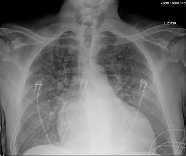
The next aspect of the diagnostic workup includes obtaining a chest radiograph (CXR). (See Figure 1.) The benefits of CXR include its wide availability, low cost, and low radiation exposure. The CXR can be used to aid in diagnosis and assess for signs of pulmonary congestion.6
Figure 1. Chest Radiograph Demonstrating Diffuse Pulmonary Edema in a Patient with Acute Decompensated Heart Failure |
 |
Source: R. Gentry Wilkerson, MD. |
A systematic review and meta-analysis by Wang et al found that a CXR showing signs of pulmonary vascular congestion significantly increased the likelihood of an HF diagnosis in patients presenting to the ED with dyspnea (likelihood ratio [LR], 12.0). They also found that the absence of cardiomegaly and the absence of pulmonary venous congestion on CXR significantly reduced the likelihood of an HF diagnosis (LR, 0.33 and 0.48, respectively). Other findings that reduce the likelihood of HF include the presence of pneumonia or hyperinflation.44 Another study by Mant et al found that any CXR finding associated with HF was 68% sensitive and 83% specific for the diagnosis.45
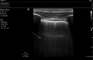
Point-of-care ultrasound (POCUS) is a bedside technique to rapidly assess for pulmonary congestion indicated by finding multiple vertical reverberation artifacts that originate from the pleural line and extend the length of the image (termed comet-tails or B-lines) due to thickening of the pulmonary interstitium because of fluid retention or fibrosis.46 The presence of two or fewer B-lines per intercostal space may be a normal finding. Three or more B-lines in an intercostal space is pathologic, with increasing numbers of B-lines corresponding to severity of pulmonary congestion.
Lung ultrasound (US) has many benefits because it is a low-cost, rapid, bedside assessment tool, and it is performed without exposure to ionizing radiation.47,48 The presence of B-lines seen on bedside ultrasound is predictive of an acute HF presentation.47 (See Figure 2.) The differential diagnosis for the presence of diffuse B-lines on lung US includes interstitial edema (both cardiogenic and noncardiogenic), acute respiratory distress syndrome (ARDS), interstitial pneumonia, and parenchymal lung disease (e.g., pulmonary fibrosis).49 Unilateral or localized pathologic B-lines suggest a localized process such as pneumonia.46
Figure 2. B Lines (arrow) on Lung Ultrasound |
 |
Source: Image used with permission from Daniel Haase, MD, RDCS, RDMS. |
Comparison studies have shown that lung US is more accurate than CXR in diagnosing pulmonary edema.50 In a 2018 systematic review by McGivery et al, the pooled sensitivity and specificity of lung US for the diagnosis of acute HF was 82.5% and 83.6%, respectively.51 A 2019 study by Pivetta et al found that lung US was more accurate than the combination of clinical examination, CXR, and natriuretic peptides.52 They also found that the use of lung US reduced diagnostic errors and reduced the median time for diagnosis from 104.5 minutes to five minutes.52 Another study by Maw et al in 2021 found that the sensitivity of lung US (88%) was superior to CXR (73%), while the specificity remained consistent (90% for both lung US and CXR).48
There are potential limitations to the use of lung US, including inter-provider reliability and specificity of findings.53 Use of lung US has a level B recommendation (reflects moderate scientific certainty) by the American College of Emergency Physicians (ACEP) in conjunction with medical history and physical examination for undifferentiated dyspneic patients with concern for acute heart failure who present to the ED.47
In addition to lung US, which provides information regarding the presence or absence of extravascular fluid, there are other forms of bedside US that may provide information regarding a patient’s volume status. Specifically, US can be used to assess for distention of the inferior vena cava (IVC) and internal jugular vein (IJV). Ultrasound also can be used to perform renal venous Doppler, which may aid in volume status assessment.54
Measurements of IVC diameter and collapsibility allow for approximation of right atrial pressure. The IVC US is performed with a phased-array or curvilinear probe in the subcostal position, with the probe oriented along the patient’s long axis. Measurement is taken 1 cm to 2 cm from the junction with the right atrium. Negative intrathoracic pressure during inspiration leads to increased RV filling and decreased IVC diameter.55
The IVC collapsibility index (also called the caval index) is calculated by dividing the difference in maximal and minimal IVC diameter by the maximal IVC diameter. [IVC collapsibility index = (IVCmax – IVCmin) / IVCmax]. Using the IVC diameter and collapsibility, one can estimate a patient’s right atrial pressure.56 An increasing IVC diameter and decreasing collapsibility correlates with increasing intravascular volume. These findings may be present even before a change in the patient’s weight or a change in symptoms.57
The IVC ultrasound may be followed during ongoing diuresis to provide serial assessments of volume status.58 During mechanical ventilation, there is increased intrathoracic pressure during inspiration leading to a reversal of the normal findings, causing an increase in IVC diameter and decreased RV filling. A distensibility index can be calculated by dividing the difference in maximal and minimal IVC diameter by the minimal IVC diameter. [IVC distensibility index = (IVCmax – IVCmin) / IVCmin].56
An elevated jugular venous pressure (JVP) is another marker for volume overload and increased right heart pressures. The clinical assessment of JVP can be difficult, especially in obese patients. Ultrasound measurement of JVD is performed using the linear probe with the patient in a semi-recumbent position (head and neck elevated to 45 degrees). The IJV diameter (JVD) is assessed within 5 cm of the jaw angle. The JVD is measured at rest and while performing a Valsalva maneuver.54
In patients without acute heart failure the JVD typically is only 0.1 cm to 0.15 cm at rest, but it can increase to approximately 1 cm during Valsalva. The JVD ratio then is calculated based on measurements at rest and during Valsalva. When intravascular congestion is present, the JVD increases at rest and the JVD ratio decreases. A JVD ratio of less than 4 is suggestive of intravascular congestion.59 (See Figure 3.)
Figure 3. Measurement of Internal Jugular Vein Diameter (JVD) in Patient with Acute Decompensated Heart Failure |
 |
Patient at rest (left, JVD = 0.52 cm) and performing Valsalva (right, JVD = 0.74 cm) shows a JVD ratio of 1.42, which is less than 4, suggestive of intravascular congestion. Source: Jonathan Hurst, MD. |
Pellicori et al evaluated the JVD ratio in patients with HF in an outpatient setting. The patients with a JVD ratio in the lower tertile had a 10-fold increased risk of death or admission for HF as compared to the patients in the upper tertile.60
Renal venous Doppler US also can be used for volume status assessment since an elevated central venous pressure (CVP) directly affects renal venous flow (RVF). The exam is performed by placing the patient in the left lateral decubitus position and identifying the right kidney in the longitudinal plane. The patient then will perform an end-expiratory hold. First, color Doppler should be used to identify an interlobar vein. Then, pulse-wave Doppler is used to assess venous flow.
Under normal conditions, RVF should be continuous, with mild variations during the cardiac cycle.61 With rising CVP, the RVF becomes discontinuous and will then form biphasic and monophasic patterns as it continues to rise.59 (See Figure 4.) Observational studies in non-hospitalized patients with HF have demonstrated that non-continuous intrakidney venous flow is associated with an increased risk of HF exacerbations and death.62,63
Figure 4. Example of Renal Venous Doppler Ultrasound in a Patient with Acute Decompensated Heart Failure |
 |
Image shows the discontinuous, monophasic pattern of renal venous flow (RVF), consistent with rising central venous pressure (CVP). Source: Jonathan Hurst, MD. |
Finally, the bedside cardiac US also will provide vital information in patients with concern for HF. The five main views in the bedside cardiac US are parasternal long axis (PLAX) view, parasternal short axis (PSAX) view, apical four chamber (A4C) view, subcostal cardiac view, and subcostal IVC view. The bedside cardiac US allows for an initial, qualitative inspection of left ventricular systolic function and estimation of EF (LVEF).
EF as a percentage is calculated by dividing the stroke volume (SV) by the end diastolic volume (EDV) of the LV and multiplying by 100 (LVEF% = SV/EDV × 100). The goal is to establish an estimated degree of ventricular dysfunction to be categorized as mild (LVEF% 45% to 54%), moderate (LVEF% 30% to 44%), or severe (LVEF% < 30%).64-66
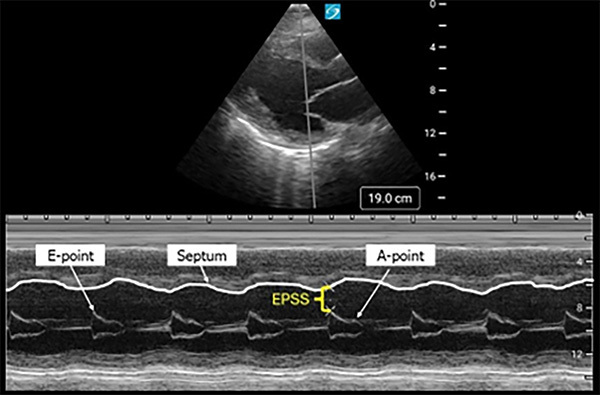
Measurement of E-point septal separation (EPSS) is another way to estimate the LVEF%. This measurement is taken in the PLAX view of the heart with the M-mode tracer over the distal tip of the anterior mitral valve leaflet (AMVL). The waveform created by movement of the AMVL creates two peaks. The larger peak is the E-point and corresponds to the maximal mitral valve opening during early diastole. The smaller peak is the A-point, which corresponds to the atrial contraction during later diastole. The distance between the E-point and the interventricular septum is the EPSS. (See Figure 5.)
Figure 5. Ultrasound Image of E-Point Septal Separation (EPSS) Taken in M-Mode |
 |
Source: Image used with permission from Youssef Annous, MD. |
Normally, there would be little separation between the E-point and the interventricular septum. Increasing EPSS is inversely associated with LVEF%. A study by McKaigney et al demonstrated that an EPSS of > 7 mm was 100% sensitive and 51.6% specific for severely reduced LVEF% (< 30%).67
The method of “eyeballing” the LVEF has been found to correlate well with more formal methods of EF measurements and may be the most useful measurement strategy overall.66 To eyeball the LVEF, one assesses the motion of the walls of the LV during systole and determines how close the AMVL gets to the septum during diastole. If the LV walls do not move much during systole and the AMVL does not approach the septum during diastole, the patient likely has a low LVEF.
Beyond the global assessment, providers should attempt to determine whether left ventricular impairment is regional or global, as a new regional wall motion abnormality is concerning for ACS.65 Additionally, bedside US is the most useful diagnostic modality for valvular dysfunction.65 Following this initial POCUS cardiac assessment, a formal echocardiogram is recommended as an inpatient for those with new HF or those with a suspected change in cardiac function, to aid in diagnosis and classification of HF.68
Laboratory Tests
The 2022 AHA/ACC/HFSA guidelines stipulate that patients diagnosed or presenting with HF should have a thorough laboratory evaluation, including complete blood count (CBC), urinalysis, basic metabolic panel (BMP), lipid profile, liver function tests, iron studies, and thyroid-stimulating hormone (TSH).3 These tests will help identify a patient’s comorbidities, aid in treatment decisions, determine etiology of HF diagnosis, and provide information regarding severity and prognosis of HF.3
For patients presenting with undifferentiated dyspnea, it is recommended to obtain either B-type natriuretic peptide (BNP) or N-terminal prohormone of B-type natriuretic peptide (NT-proBNP) to support or exclude a diagnosis of HF.3 The half-life of BNP is 20 minutes and the half-life of NT-proBNP is 60-120 minutes.69 Use of BNP to diagnose acute HF was established in the Breathing Not Properly (BNP) study. This study demonstrated that using a cutoff of 100 picograms (pg) per milliliter (mL), the diagnostic accuracy was 83.4%. Using a cutoff of < 50 pg/mL, the negative predictive value was 96%.70
The N-Terminal Pro-BNP Investigation of Dyspnea in the Emergency Department (PRIDE) Study established the use of NT-proBNP for the diagnosis of acute HF. In this study, an NT-proBNP level < 300 pg/mL had a 99% negative predictive value for acute HF. For patients younger than 50 years of age, a cutoff of 450 pg/mL had a sensitivity of 93% and specificity of 95%. The positive predictive value was 67%. For patients older than 50 years of age, use of 900 pg/mL as a cutoff was associated with a 91% sensitivity, specificity of 80%, and a positive predictive value of 77%.71
The use of BNP or NT-proBNP is interchangeable and dependent on the test available at the treating institution. Levels of BNP have been found to increase in those with left ventricular dysfunction, correlate with a patient’s NYHA class, and provide prognostic value.70,72 The 2021 ESC guidelines recommend cutoffs of < 35 pg/mL for BNP and < 125 pg/mL to rule out HF in the non-acute setting.25
Atrial natriuretic peptide (ANP) also is released in response to stretching of the atrial wall. The half-life of ANP is only two minutes and, therefore, it is less useful for diagnostic purposes.69 In the emergency setting, natriuretic peptides have greater sensitivity than specificity. In other words, they are more useful in ruling out HF than ruling it in.3 Of note, obesity is associated with lower levels of BNP and NT-proBNP, which reduces their overall sensitivity.73 Other factors, including age, sex, and comorbidities, such as renal dysfunction and arrhythmias, may influence natriuretic peptide levels.70,74
Management
The management strategy used should be tailored to the specific HF syndrome with which the patient presents. Key aspects to consider with initial management decisions include the patient’s hemodynamics and volume status. Management options for the special population of patients with cardiogenic shock follow the sections on medication classes. (See Table 4.)
Table 4. Medications Used in the Treatment of Acute Heart Failure |
|||
Medication |
Mechanism |
Dosing |
Comment |
Diuretics |
|||
Furosemide Torsemide Bumetanide |
Loop diuretic |
No prior diuretic use:107
Prior diuretic use:76
|
“40-20-1 Rule”: 40 mg furosemide = 20 mg torsemide = 1 mg bumetanide May augment diuresis with sequential nephron blockade by adding thiazide diuretic108
|
Vasodilators |
|||
Nitroglycerin |
Nitrate |
Initial dose: 5 mcg to 10 mcg/min IV, increase by 10 mcg to 20 mcg/min q3-5 min109
|
Typical dose: 100 mcg/min to 200 mcg/min Also may start with 0.4 mg SL q5 min while waiting on IV access |
Nitroprusside |
Vasodilator |
Initial dose: 5 mcg/min to 10 mcg/min, increase q5 min to 5 mcg/min to 400 mcg/min109
|
Monitor for cyanide toxicity, typically dose- and duration-dependent Risk of rebound vasoconstriction after discontinuation89 |
Captopril |
ACE Inhibitor |
12.5 mg to 25 mg SL91 |
SL captopril may be less desirable due to need to remove BiPAP mask for administration |
Enalaprilat |
ACE Inhibitor |
1.25 mg IV q15 min92 |
|
Vasopressors |
|||
Norepinephrine (NE) |
Adrenergic agonist |
Initial dose: 0.02 mcg/kg/min, titrate up to maintain MAP goal108 |
NE is the first-line vasopressor in treatment of cardiogenic shock Use of vasopressors increases myocardial oxygen, increases risk of arrhythmias |
Epinephrine |
Adrenergic agonist |
Initial dose: 0.01 mcg/kg/min to 0.05 mcg/kg/min108
|
|
Inotropes |
|||
Dobutamine |
Adrenergic agonist |
Initial dose: 2 mcg/kg/min to 5 mcg/kg/min108
|
Associated with an increased risk of ventricular arrhythmias May cause hypotension at high doses due to peripheral vasodilation |
Milrinone |
Phosphodies-terase inhibitor |
Initial dose: 0.125 mcg/kg/min to 0.25 mcg/kg/min108
|
Associated with an increased risk of ventricular arrhythmias Patients on beta-blockers may have greater benefit from milrinone than dobutamine |
Adjunct |
|||
Tolvaptan |
Vasopressin antagonist |
15 mg daily110
|
Requires close monitoring of serum sodium levels |
PO: orally; IV: intravenous; HTN: hypertension; SL: sublingual; ACE: angiotensin converting enzyme; BiPAP: bilevel positive airway pressure; MAP: mean arterial pressure |
|||
Diuretics
For the patients presenting with ADHF, the focus of therapy should be on decongestion.2 The recommended therapy when initiating diuresis is with intravenous (IV) loop diuretics.2 The use of loop diuretics will inhibit water and sodium reabsorption and therefore increase excretion of chloride, calcium, and magnesium. This mechanism helps to reduce preload.47,75 Commonly used loop diuretics include furosemide, torsemide, and bumetanide. When given intravenously, these medications differ in potency, with furosemide being the least potent, followed by torsemide and bumetanide.47
When using IV loop diuretics, it is important to consider potential side effects. Common adverse effects include the development of intravascular hypovolemia, electrolyte disturbances (typically sodium, potassium, calcium, and magnesium), and worsening renal function.75 Worsening renal function may be related to activation of the renin-aldosterone-angiotensin and sympathetic nervous systems.76 Diuresis with loop diuretics may be augmented with the concomitant use of thiazide diuretics.3
IV furosemide is expected to have a quick diuretic effect (within 30 minutes), with peak effect occurring at about 1.5 hours.76 In terms of dosing strategy, there is uncertainty regarding dosing recommendations. Initial IV furosemide dose recommendations range from 100% to 250% of the patient’s home oral dose.77 Higher doses are required for those presenting with decreased renal function. For example, a patient taking a home oral dose of 40 mg furosemide daily would be given 80 mg IV furosemide to treat an acute HF presentation if following a recommendation to give 200% of the home dose.
Torsemide is another diuretic that has a longer half-life and increased potency compared to furosemide.78 The TOrasemide In Congestive Heart Failure (TORIC) study was an open-label non-randomized study that compared the daily use of torsemide to furosemide and other diuretics for chronic HF. There was a > 50% decrease in mortality at the end of the study for patients taking torsemide as compared to furosemide and other diuretics (2.2% vs. 4.5%; P < 0.05). It also was shown that the use of torsemide was associated with a higher likelihood of improving NYHA class by at least one level and a 10-fold lower rate of needing potassium supplementation.79
Earlier administration of diuretics is recommended in those patients suspected of having heart failure exacerbation.47 Specific timing for diuretic administration is not known. A study using data from the ADHERE-EM registry of encounters for patients ≥ 65 years of age for acute heart failure who received IV HF medications by Wong et al showed that there was no difference due to time to diuretic in the composite outcome of 30-day all-cause mortality and hospital readmission (HR, 1.00).80 However, they did find that there was a minimally increased risk of in-hospital mortality with every hour increase in the time to treatment (adjusted odds ratio [aOR], 1.01).80
Using more recent data from the REPORT-HF registry, Ouwerkerk et al found that diuretic use within 30 minutes of hospital arrival compared to diuretic use 12-24 hours after arrival did not result in a statistically significant rate of in-hospital mortality. However, for those who survived to hospital discharge, there was a statistical difference in 30-day all-cause mortality for patients who received IV diuretics within 30 minutes compared to those who received IV diuretics six to 12 hours after presentation (2.7% vs. 4.1%). Thirty-day mortality for patients who received IV diuretics 12-24 hours after presentation was 2.2%, the lowest of all the groups. Diuresis goals include net fluid loss and decrease in weight by at least 1 kg/day.81
Nitrates
Patients with ADHF experience both excess preload and afterload, which contributes to their clinical presentation. Vasodilators are recommended to reduce preload and afterload, allowing for improvement in cardiac output and reduced pulmonary edema.47 Nitrates are the vasodilator of choice for treatment of ADHF in patients who are hypertensive or normotensive.82,83 Nitrates have multiple favorable properties, including their effect on both preload and afterload, quick onset and half-life, and overall safety. Nitrates have been shown to have a primary effect of decreasing preload but will also reduce afterload at higher doses.47
Several studies have shown the efficacy and safety of high-dose nitrates in the treatment of ADHF. A study by Levy et al evaluated the use of high-dose IV nitroglycerin in hypertensive patients presenting with ADHF who failed initial therapies. They found reduced rates of noninvasive positive pressure ventilation use, intubation, and ICU admission in patients receiving high-dose nitroglycerin.84 The HI-DOSE SCAPE study described the characteristics of patients who received nitroglycerin infusions of > 100 mcg/min for the management of H-AHF at a single academic institution. Lack of a control group limits the interpretation of the findings, but a feared complication of high-dose nitroglycerin treatment, hypotension, was only seen in 4% of patients.85 The recent ACEP guidelines for HF have a level C recommendation (consensus) for the use of high-dose nitroglycerin in patients with ADHF presenting to the ED.47
The use of IV isosorbide dinitrate also has been shown to have clinical benefits in the treatment of acute HF. In a study by Cotter et al, patients presenting to the ED with signs and symptoms of congestive heart failure were randomized to receive either high-dose isosorbide dinitrate plus low-dose furosemide, or low-dose isosorbide dinitrate with high-dose furosemide. They found that patients in the high-dose nitrate group had significantly fewer occurrences of the composite endpoint of intubation, myocardial infarction, or death (25% vs. 46%; P = 0.041).86 The prolonged half-life of four hours vs. three to five minutes for IV nitroglycerin makes IV isosorbide dinitrate a less appealing option in the event of hypotension. Caution should be given to those patients who arrive hypotensive, bradycardic, or report recent use of phosphodiesterase type-5 inhibitors.
Alternative Agents
Sodium nitroprusside is a favorable option in patients presenting with ADHF, especially for those who are significantly hypertensive. Similarly to nitrates, nitroprusside provides both venous and arterial dilation, providing treatment of both elevated preload and afterload.87 It has the advantage of a very short half-life, allowing for rapid titration. A study by Garatti et al found that the use of sodium nitroprusside for patients presenting with acute heart failure was safe and effective, especially for patients with a dilated LV.88 Potential drawbacks of nitroprusside include the need for close hemodynamic monitoring because of the concern for hypotension, risk of cyanide toxicity with prolonged use, and risk for rebound vasoconstriction after the infusion is stopped.89
Angiotensin converting-enzyme (ACE) inhibitors are another vasodilatory option for the treatment of ADHF. ACE inhibitors provide reductions in both preload and afterload. In addition, ACE inhibitors can combat the high renin state that is common in these patients.90 Commonly used formulations include sublingual captopril or IV enalaprilat. In a trial by Hamilton et al, patients presenting with acute pulmonary edema were evaluated after receiving a dose of sublingual captopril vs. placebo, following standard treatment of oxygen, nitrates, morphine, and furosemide. Patients who received sublingual captopril were shown to have more rapid clinical improvement than the standard regimen alone.91
Studies looking at the use of IV enalapril also have shown improvements in acute heart failure management. A retrospective cohort study by Ayaz et al showed that a single dose of IV enalaprilat led to significant reductions in SBP (-30.5 mmHg), with low rates of hypotension (1.9%), and no significant change in renal function.92 A key drawback of these ACE therapies is that they are not titratable, increasing the risk for patients to develop hypotension.
The use of vasopressin antagonists (tolvaptan) has been investigated as a potential adjunct therapy for the acute treatment of heart failure. The EVEREST trial was a randomized trial in which patients hospitalized for HF were given either oral tolvaptan or placebo, in addition to standard therapy. Patients who received tolvaptan had improvement in reported dyspnea and body weight, but no difference in mortality (P = 0.68) or a composite measure of cardiovascular death and readmission for HF (P = 0.55).93 Hyponatremic patients in the tolvaptan group did show improvement in serum sodium levels.93 Therefore, tolvaptan can be considered as an adjunct for acute heart failure management in patients who are volume-overloaded and hyponatremic.94
Noninvasive Ventilation
Noninvasive ventilation (NIV), including continuous positive airway pressure (CPAP) and bilevel positive airway pressure (BiPAP), is considered first-line therapy in patients with ADHF with pulmonary edema.95 NIV provides positive pressure, which has the benefit of increasing intrathoracic pressure. This causes decreased venous return, thus reducing right- and left-sided preload.25 Positive airway pressure increases oxygenation and provides ventilatory support, decreasing the work of breathing and improving hypercapnia.25,96,97
NIV has been shown to decrease rates of endotracheal intubation and even mortality in high-risk patients.95,98 The 3CPO trial was a multicenter, randomized controlled trial that compared NIV (CPAP and BiPAP) vs. conventional oxygen therapy in patients presenting with acute cardiogenic pulmonary edema. They found no difference in mortality between groups but did find that NIV leads to more rapid improvements in dyspnea, heart rate, acidosis, and hypercapnia.99
In addition, evidence supports the use of NIV in the prehospital setting for patients suspected of having ADHF with pulmonary edema. A trial by Ducros et al looked at these patients in the prehospital setting and compared standard treatment to standard treatment with the addition of CPAP. They found significant improvements in subjective symptoms, vital signs, and rates of intubation in patients treated with CPAP.100
When using CPAP, it is recommended to start at 5 cmH2O and titrate up as needed. For BiPAP, it is recommended to start at 10/5 cmH2O and titrate up as needed, ensuring that expired tidal volumes are greater than 4 mL/kg to 6 mL/kg.95 The latest ESC 2021 guidelines recommend the use of NIV both in the prehospital and ED in patients with acute heart failure and respiratory distress. Their usage is recommended “to improve gas exchange and reduce the rate of endotracheal intubation.”25 If respiratory failure continues to progress despite the use of oxygen or NIV, intubation is recommended.25
Cardiogenic Shock
Cardiogenic shock is a high-risk situation with significant morbidity and mortality. Cardiogenic shock is characterized by hypotension with evidence of end-organ perfusion. Given that the most common etiology of cardiogenic shock is acute myocardial infarction (accounting for an estimated 80% of cases), the most important treatment strategy is early revascularization.101,102 In the SHOCK trial, patients with evidence of shock in the setting of myocardial infarction were randomized to either emergency revascularization or initial medical stabilization. Although there was no difference in the primary end point of 30-day mortality (46.7% vs. 56.0%; P = 0.11), there was a significant improvement in six-month mortality in the early revascularization group (50.3% vs. 63.1%; P = 0.027).102
Aside from revascularization, additional management considerations in cardiogenic shock include IV fluids, vasopressors, and inotropes.101 Initial attempts at resuscitation toward improvement in end-organ perfusion may include careful administration of small fluid boluses in patients who do not show signs of acute pulmonary edema or right ventricular overload.103 For patients who continue to be hypotensive, norepinephrine is the recommended first vasopressor used in treatment of cardiogenic shock.101,103
Vasopressors do carry the risk of increasing myocardial oxygen demand and increasing the risk of arrhythmias. Increasing afterload from rising vasopressor doses may cause worsening myocardial function. Therefore, it is recommended that vasopressors be used for the shortest possible duration and lowest dose.101
Inotropes, such as dobutamine or milrinone, are seen as a temporizing measure for patients with severely reduced cardiac function. It is recommended that inotropes be used in cardiogenic shock for maintenance of systemic perfusion until more definitive therapies can be provided.94 Inotropes have the potentially harmful effects of increasing myocardial ischemia and causing arrhythmias.104 The OPTIME-CHF trial showed that the routine use of milrinone infusion in patients presenting with an HF exacerbation led to significant increases in hypotension and arrhythmias, with a nonsignificant increase in mortality.104
Dobutamine acts primarily through stimulation of beta-1 adrenergic receptors, causing increased stroke volume and cardiac output, in addition to decreased systemic vascular resistance.105 Milrinone is a phosphodiesterase inhibitor that facilitates vascular smooth muscle relaxation. This provides increased cardiac output with reduced left ventricular afterload.106
If signs of end-organ perfusion continue after initial resuscitation, patients may be considered for mechanical circulatory support. Options for mechanical support include IABP, left-ventricular assist devices (LVAD), or extracorporeal membrane oxygenation (ECMO).101 Decisions regarding mechanical support should be made in conjunction with cardiology and cardiothoracic surgery teams.
Disposition
New onset or acute decompensated heart failure is associated with a high admission rate of more than 80%.111 This likely is related to the need for complex outpatient care and morbidity and mortality associated with these presentations. Although HF does present a high-risk scenario, several studies have shown that there is potential for discharging more patients safely to home. Determining if there are certain decision-making tools that can allow ED providers to identify low-risk patients and safely discharge them home remains an important topic of discussion.
The Ottawa Heart Failure Risk Score (OHFRS) score has a Level B recommendation from the recent ACEP guidelines to help identify patients with high-risk features who should not be discharged home.47 The OHFRS score was prospectively evaluated by Stiell et al in adult patients presenting to Canadian EDs with signs and symptoms consistent with acute heart failure.112 The score looked to identify serious adverse events (SAEs) within 14 days of disposition. The OHFRS score considers various aspects of the patient’s history, initial vital signs, laboratory and ECG findings, and vital signs during a six-minute walk test to help categorize patients into one of four risk categories. Scores range from 0 to 15, with higher scores being associated with increased risk of 14-day SAEs. The OHFRS score does not appear either sensitive or specific enough to be considered the sole criterion to determine disposition from the ED.47 However, the score showed the ability to perform better than the standard physician’s assessment in determining which patients should not be discharged home due to the presence of high-risk features.
The complexities of home healthcare and outpatient management for patients with HF also contribute to the high admission rate. When considering discharge, ED providers should consider several social factors to aid their decision-making. These include the patient’s ability to follow up as an outpatient, understanding of return precautions, compliance with medical therapy, and ability to monitor weight at home.113 If discharge is determined to be reasonable, patients should have close follow-up arranged with either their primary care physician or cardiologist.
Given the risks associated with acute heart failure, complexities of medical therapy, and multiple patient factors to be considered at discharge, it is reasonable to suggest that most HF patients should continue to be admitted. Additional research is necessary to help identify those patients at high risk and determine whether a patient can be safely cared for as an outpatient.
References
- Bozkurt B, Coats AJ, Tsutsui H, et al. Universal Definition and Classification of Heart Failure: A Report of the Heart Failure Society of America, Heart Failure Association of the European Society of Cardiology, Japanese Heart Failure Society and Writing Committee of the Universal Definition of Heart Failure. J Card Fail 2021;27:S1071-9164(21)00050-6. [Online ahead of print].
- Mitter SS, Pinney SP. Advances in the management of acute decompensated heart failure. Med Clin North Am 2020;104:601-614.
- Heidenreich PA, Bozkurt B, Aguilar D, et al. 2022 AHA/ACC/HFSA Guideline for the Management of Heart Failure: A Report of the American College of Cardiology/American Heart Association Joint Committee on Clinical Practice Guidelines. Circulation 2022;145:e895-e1032.
- Virani SS, Alonso A, Benjamin EJ, et al. Heart disease and stroke statistics—2020 update: A report from the American Heart Association. Circulation 2020;141:e139-e596.
- Heidenreich PA, Albert NM, Allen LA, et al. Forecasting the impact of heart failure in the United States: A policy statement from the American Heart Association. Circ Heart Fail 2013;6:606-619.
- Shahim B, Kapelios CJ, Savarese G, Lund LH. Global public health burden of heart failure: An updated review. Card Fail Rev 2023;9:e11.
- Lan T, Liao YH, Zhang J, et al. Mortality and readmission rates after heart failure: A systematic review and meta-analysis. Ther Clin Risk Manag 2021;17:1307-1320.
- Zhang X, Qiu P, Prushinskaya A, et al. Characteristics of emergency department admissions with congestive heart failure in the United States: A nationwide cross-sectional study. BMC Emerg Med 2022;22:16.
- Fonarow GC, Abraham WT, Albert NM, et al. Factors identified as precipitating hospital admissions for heart failure and clinical outcomes: Findings from OPTIMIZE-HF. Arch Intern Med 2008;168:847-854.
- Arrigo M, Gayat E, Parenica J, et al; GREAT Network. Precipitating factors and 90-day outcome of acute heart failure: A report from the intercontinental GREAT registry. Eur J Heart Fail 2017;19:201-208.
- Kurmani S, Squire I. Acute heart failure: Definition, classification and epidemiology. Curr Heart Fail Rep 2017;14:385-392.
- Hummel A, Empen K, Dörr M, Felix SB. De novo acute heart failure and acutely decompensated chronic heart failure. Dtsch Arztebl Int 2015;112:298-310.
- Raffaello WM, Henrina J, Huang I, et al. Clinical characteristics of de novo heart failure and acute decompensated chronic heart failure: Are they distinctive phenotypes that contribute to different outcomes? Card Fail Rev 2021;7:e02.
- Pranata R, Tondas AE, Yonas E, et al. Differences in clinical characteristics and outcome of de novo heart failure compared to acutely decompensated chronic heart failure – systematic review and meta-analysis. Acta Cardiol 2021;76:410-420.
- Xanthopoulos A, Butler J, Parissis J, et al. Acutely decompensated versus acute heart failure: Two different entities. Heart Fail Rev 2020;25:907-916.
- Younis A, Mulla W, Goldkorn R, et al. Differences in mortality of new-onset (de-novo) acute heart failure versus acute decompensated chronic heart failure. Am J Cardiol 2019;124:554-559.
- Harjola VP, Follath F, Nieminen MS, et al. Characteristics, outcomes, and predictors of mortality at 3 months and 1 year in patients hospitalized for acute heart failure. Eur J Heart Fail 2010;12:239-248.
- Sliwa K, Hilfiker-Kleiner D, Petrie MC, et al. Current state of knowledge on aetiology, diagnosis, management, and therapy of peripartum cardiomyopathy: A position statement from the Heart Failure Association of the European Society of Cardiology Working Group on peripartum cardiomyopathy. Eur J Heart Fail 2010;12:767-778.
- Bhattacharyya A, Basra SS, Sen P, Kar B. Peripartum cardiomyopathy: A review. Tex Heart Inst J 2012;39:8-16.
- Bauersachs J, König T, van der Meer P, et al. Pathophysiology, diagnosis and management of peripartum cardiomyopathy: A position statement from the Heart Failure Association of the European Society of Cardiology Study Group on peripartum cardiomyopathy. Eur J Heart Fail 2019;21:827-843.
- Iorgoveanu C, Zaghloul A, Ashwath M. Peripartum cardiomyopathy: A review. Heart Fail Rev 2021;26:1287-1296.
- Halpern DG, Weinberg CR, Pinnelas R, et al. Use of medication for cardiovascular disease during pregnancy: JACC state-of-the-art review. J Am Coll Cardiol 2019;73:457-576.
- Forrester JS, Diamond G, Chatterjee K, Swan HJ. Medical therapy of acute myocardial infarction by application of hemodynamic subsets. N Engl J Med 1976;295:1404-1413.
- Nohria A, Tsang SW, Fang JC, et al. Clinical assessment identifies hemodynamic profiles that predict outcomes in patients admitted with heart failure. J Am Coll Cardiol 2003;41:1797-1804.
- McDonagh TA, Metra M, Adamo M, et al. 2021 ESC guidelines for the diagnosis and treatment of acute and chronic heart failure: Developed by the task force for the diagnosis and treatment of acute and chronic heart failure of the European Society of Cardiology (ESC). With the special contribution of the Heart Failure Association (HFA) of the ESC. Eur J Heart Fail 2022;24:4-131.
- Nieminen MS, Böhm M, Cowie MR, et al. Executive summary of the guidelines on the diagnosis and treatment of acute heart failure: The Task Force on Acute Heart Failure of the European Society of Cardiology. Eur Heart J 2005;26:384-416.
- Nieminen MS, Brutsaert D, Dickstein K, et al. EuroHeart Failure Survey II (EHFS II): A survey on hospitalized acute heart failure patients: Description of population. Eur Heart J 2006;27:2725-2736.
- Dickstein K, Cohen-Solal A, Filippatos G, et al. ESC guidelines for the diagnosis and treatment of acute and chronic heart failure 2008. The Task Force for the Diagnosis and Treatment of Acute and Chronic Heart Failure 2008 of the European Society of Cardiology. Developed in collaboration with the Heart Failure Association of the ESC (HFA) and endorsed by the European Society of Intensive Care Medicine (ESICM). Eur J Heart Fail 2008;10:933-989.
- Wang K, Samai K. Role of high-dose intravenous nitrates in hypertensive acute heart failure. Am J Emerg Med 2020;38:132-137.
- Paone S, Clarkson L, Sin B, Punnapuzha S. Recognition of sympathetic crashing acute pulmonary edema (SCAPE) and use of high-dose nitroglycerin infusion. Am J Emerg Med 2018;36:1526.e5-1526.e7.
- Mathew R, Kumar A, Sahu A, et al. High-dose nitroglycerin bolus for sympathetic crashing acute pulmonary edema: A prospective observational pilot study. J Emerg Med 2021;61:271-277.
- Vahdatpour C, Collins D, Goldberg S. Cardiogenic shock. J Am Heart Assoc 2019;8:e011991.
- Harjola VP, Lassus J, Sionis A, et al. Clinical picture and risk prediction of short-term mortality in cardiogenic shock. Eur J Heart Fail 2015;17:501-509.
- Hochman JS, Sleeper LA, Godfrey E, et al. SHould we emergently revascularize Occluded Coronaries for cardiogenic shocK: An international randomized trial of emergency PTCA/CABG-trial design. Am Heart J 1999;137:313-321.
- Thiele H, Zeymer U, Neumann FJ, et al. Intraaortic balloon support for myocardial infarction with cardiogenic shock. N Engl J Med 2012;367:1287-1296.
- Baran DA, Grines CL, Bailey S, et al. SCAI clinical expert consensus statement on the classification of cardiogenic shock. Catheter Cardiovasc Interv 2019;9429-9437.
- Kostura M, Smalley C, Koyfman A, Long B. Right heart failure: A narrative review for emergency clinicians. Am J Emerg Med 2022;58:106-113.
- Logeart D, Isnard R, Resche-Rigon M, et al. Current aspects of the spectrum of acute heart failure syndromes in a real-life setting: The OFICA study. Eur J Heart Fail 2013;15465-476.
- Thandavarayan RA, Chitturi KR, Guha A. Pathophysiology of acute and chronic right heart failure. Cardiol Clin 2020;38:149-160.
- Brown SB, Raina A, Katz D, et al. Longitudinal shortening accounts for the majority of right ventricular contraction and improves after pulmonary vasodilator therapy in normal subjects and patients with pulmonary arterial hypertension. Chest 2011;140:27-33.
- Sheehan F, Redington A. The right ventricle: Anatomy, physiology and clinical imaging. Heart 2008;94:1510-1515.
- Mehta PA, Dubrey SW. High output heart failure. QJM 2009;102:235-241.
- de Ávila DX, Villacorta H, de Andrade Martins W, Mesquita ET. High-output cardiac failure: A forgotten phenotype in clinical practice. Curr Cardiol Rev 2022;18:e050821195319.
- Wang CS, FitzGerald JM, Schulzer M, et al. Does this dyspneic patient in the emergency department have congestive heart failure? JAMA 2005;294:1944-1956.
- Mant J, Doust J, Roalfe A, et al. Systematic review and individual patient data meta-analysis of diagnosis of heart failure, with modelling of implications of different diagnostic strategies in primary care. Health Technol Assess 2009;13:1-207.
- Marini TJ, Rubens DJ, Zhao YT, et al. Lung ultrasound: The essentials. Radiol Cardiothorac Imaging 2021;3:e200564.
- Silvers SM, Howell JM, Kosowsky JM, et al. Clinical policy: Critical issues in the evaluation and management of adult patients presenting to the emergency department with acute heart failure syndromes. Ann Emerg Med 2007;49:627-669.
- Maw AM, Hassanin A, Ho PM, et al. Diagnostic accuracy of point-of-care lung ultrasonography and chest radiography in adults with symptoms suggestive of acute decompensated heart failure: A systematic review and meta-analysis. JAMA Netw Open 2019;2:e190703.
- Volpicelli G, Elbarbary M, Blaivas M, et al. International evidence-based recommendations for point-of-care lung ultrasound. Intensive Care Med 2012;38:577-591.
- Martindale JL, Noble VE, Liteplo A. Diagnosing pulmonary edema: Lung ultrasound versus chest radiography. Eur J Emerg Med 2013;20:356-360.
- McGivery K, Atkinson P, Lewis D, et al. Emergency department ultrasound for the detection of B-lines in the early diagnosis of acute decompensated heart failure: A systematic review and meta-analysis. CJEM 2018;20:343-352.
- Pivetta E, Goffi A, Nazerian P, et al. Lung ultrasound integrated with clinical assessment for the diagnosis of acute decompensated heart failure in the emergency department: A randomized controlled trial. Eur J Heart Fail 2019;21:754-766.
- Picano E, Scali MC, Ciampi Q, Lichtenstein D. Lung ultrasound for the cardiologist. JACC Cardiovasc Imaging 2018;11:1692-1705.
- Pellicori P, Platz E, Dauw J, et al. Ultrasound imaging of congestion in heart failure: Examinations beyond the heart. Eur J Heart Fail 2021;23:703-712.
- Lang RM, Badano LP, Victor MA, et al. Recommendations for cardiac chamber quantification by echocardiography in adults: An update from the American Society of Echocardiography and the European Association of Cardiovascular Imaging. J Am Soc Echocardiogr 2015;28:1-39.
- Kaptein MJ, Kaptein EM. Inferior vena cava collapsibility index: Clinical validation and application for assessment of relative intravascular volume. Adv Chronic Kidney Dis 2021;28:218-226.
- Dovancescu S, Pellicori P, Mabote T, et al. The effects of short-term omission of daily medication on the pathophysiology of heart failure. Eur J Heart Fail 2017;19:643-649.
- Laffin LJ, Patel AV, Saha N, et al. Focused cardiac ultrasound as a predictor of readmission in acute decompensated heart failure. Int J Cardiovasc Imaging 2018;34:1075-1079.
- Pellicori P, Shah P, Cuthbert J, et al. Prevalence, pattern and clinical relevance of ultrasound indices of congestion in outpatients with heart failure. Eur J Heart Fail 2019;21:904-916.
- Pellicori P, Kallvikbacka-Bennett A, Dierckx R, et al. Prognostic significance of ultrasound-assessed jugular vein distensibility in heart failure. Heart 2015;101:1149-158.
- Jeong SH, Jung DC, Kim SH, Kim SH. Renal venous doppler ultrasonography in normal subjects and patients with diabetic nephropathy: Value of venous impedance index measurements. J Clin Ultrasound 2011;39:512-518.
- Puzzovivo A, Monitillo F, Guida P, et al. Renal venous pattern: A new parameter for predicting prognosis in heart failure outpatients. J Cardiovasc Dev Dis 2018;5:52.
- Husain-Syed F, Singam NSV, Viehman JK, et al. Changes in Doppler-derived kidney venous flow and adverse cardiorenal outcomes in patients with heart failure. J Am Heart Assoc 2023;12:e030145.
- Ruben M, Molinas MS, Paladini H, et al. Emerging concepts in heart failure management and treatment: Focus on point-of-care ultrasound in cardiogenic shock. Drugs Context 2023;12:2022-5-8.
- Bansal M, Ranjan S. Echocardiography in the management of cardiogenic shock. Indian J Clin Cardiol 2020;1:20-30.
- Gudmundsson P, Rydberg E, Winter R, Willenheimer R. Visually estimated left ventricular ejection fraction by echocardiography is closely correlated with formal quantitative methods. Int J Cardiol 2005;101:209-212.
- McKaigney CJ, Krantz MJ, La Rocque CL, et al. E-point septal separation: A bedside tool for emergency physician assessment of left ventricular ejection fraction. Am J Emerg Med 2014;32:493-497.
- Heart Failure Society of America; Lindenfeld J. Albert NM, Boehmer JP, et al. HFSA 2010 Comprehensive Heart Failure Practice Guideline. J Card Fail 2010;16:e1-e194.
- Baba M, Yoshida K, Ieda M. Clinical applications of natriuretic peptides in heart failure and atrial fibrillation. Int J Mol Sci 2019;20:2824.
- Maisel AS, Krishnaswamy P, Nowak RM, et al. Rapid measurement of B-type natriuretic peptide in the emergency diagnosis of heart failure. N Engl J Med 2002;347:161-167.
- Januzzi JL Jr, Camargo CA, Anwaruddin S, et al. The N-terminal Pro-BNP investigation of dyspnea in the emergency department (PRIDE) study. Am J Cardiol 2005;95:948-854.
- Harrison A, Morrison LK, Krishnaswamy P, et al. B-Type natriuretic peptide predicts future cardiac events in patients presenting to the emergency department with dyspnea. Ann Emerg Med 2002;39:131-138.
- Horwich TB, Hamilton MA, Fonarow GC. B-type natriuretic peptide levels in obese patients with advanced heart failure. J Am Coll Cardiol 2006;47:85-90.
- Tang WHW, Girod JP, Lee MJ, et al. Plasma B-type natriuretic peptide levels in ambulatory patients with established chronic symptomatic systolic heart failure. Circulation 2003;108:2964-2966.
- Wu MY, Chang NC, Su CL, et al. Loop diuretic strategies in patients with acute decompensated heart failure: A meta-analysis of randomized controlled trials. J Crit Care 2014;29:2-9.
- Felker GM, O’Connor CM, Braunwald E. Loop diuretics in acute decompensated heart failure: Necessary? Evil? A necessary evil? Circ Heart Fail 2009;2:56-62.
- Felker GM, Lee KL, Bull DA, et al. Diuretic strategies in patients with acute decompensated heart failure. N Engl J Med 2011;364:797-805.
- Suri SS, Pamboukian SV. Optimal diuretic strategies in heart failure. Ann Transl Med 2021;9:517.
- Cosín J, Díez J; TORIC investigators. Torasemide in chronic heart failure: Results of the TORIC study. Eur J Heart Fail 2002;4:507-513.
- Wong YW, Fonarow GC, Mi X, et al. Early intravenous heart failure therapy and outcomes among older patients hospitalized for acute decompensated heart failure: Findings from the Acute Decompensated Heart Failure Registry Emergency Module (ADHERE-EM). Am Heart J 2013;166:349-356.
- Hollenberg SM, Warner Stevenson L, Ahmad T, et al. 2019 ACC expert consensus decision pathway on risk assessment, management, and clinical trajectory of patients hospitalized with heart failure: A report of the American College of Cardiology Solution Set Oversight Committee. J Am Coll Cardiol 2019;74:1966-2011.
- Collins SP, Levy PD, Martindale JL, et al. Clinical and research considerations for patients with hypertensive acute heart failure: A Consensus Statement from the Society for Academic Emergency Medicine and the Heart Failure Society of America Acute Heart Failure Working Group. Acad Emerg Med 2016;23:922-931.
- Mebazaa A, Yilmaz MB, Levy P, et al. Recommendations on pre-hospital and early hospital management of acute heart failure: A consensus paper from the Heart Failure Association of the European Society of Cardiology, the European Society of Emergency Medicine and the Society of Academic Emergency Medicine - short version. Eur Heart J 2015;36:1958-1966.
- Levy P, Compton S, Welch R, et al. Treatment of severe decompensated heart failure with high-dose intravenous nitroglycerin: A feasibility and outcome analysis. Ann Emerg Med 2007;50:144-152.
- Houseman BS, Martinelli AN, Oliver WD, et al. High-dose nitroglycerin infusion description of safety and efficacy in sympathetic crashing acute pulmonary edema: The HI-DOSE SCAPE study. Am J Emerg Med 2023;63:74-78.
- Cotter G, Metzkor E, Kaluski E, et al. Randomised trial of high-dose isosorbide dinitrate plus low-dose furosemide versus high-dose furosemide plus low-dose isosorbide dinitrate in severe pulmonary oedema. Lancet 1998;351:389-393.
- Opasich C, Cioffi G, Gualco A. Nitroprusside in decompensated heart failure: What should a clinician really know? Curr Heart Fail Rep 2009;6:182-190.
- Garatti L, Frea S, Bocchino PP, et al. Sodium nitroprusside in acute heart failure: A multicenter historic cohort study. Int J Cardiol 2022;369:37-44.
- Packer M, Meller J, Medina N, et al. Rebound hemodynamic events after the abrupt withdrawal of nitroprusside in patients with severe chronic heart failure. N Engl J Med 1979;301:1193-1197.
- Packer M, Lee WH, Medina N, et al. Functional renal insufficiency during long-term therapy with captopril and enalapril in severe chronic heart failure. Ann Intern Med 1987;106:346-354.
- Hamilton RJ, Carter WA, Gallagher EJ. Rapid improvement of acute pulmonary edema with sublingual captopril. Acad Emerg Med 1996;3:205-212.
- Ayaz SI, Sharkey CM, Kwiatkowski GM, et al. Intravenous enalaprilat for treatment of acute hypertensive heart failure in the emergency department. Int J Emerg Med 2016;9:28.
- Konstam MA, Gheorghiade M, Burnett JC Jr, et al. Effects of oral tolvaptan in patients hospitalized for worsening heart failure: The EVEREST outcome trial. JAMA 2007;297:1319-1331.
- Yancy CW, Jessup M, Bozkurt B, et al. 2013 ACCF/AHA guideline for the management of heart failure: Executive summary: A report of the American College of Cardiology Foundation/American Heart Association Task Force on Practice Guidelines. Circulation 2013;128:1810-1852.
- Masip J. Noninvasive ventilation in acute heart failure. Curr Heart Fail Rep 2019;16:89-97.
- Tobin MJ. Advances in mechanical ventilation. N Engl J Med 2001;344:1986-1996.
- Tallman TA, Peacock WF, Emerman CL, et al; ADHERE Registry. Noninvasive ventilation outcomes in 2,430 acute decompensated heart failure patients: An ADHERE registry analysis. Acad Emerg Med 2008;15:355-362.
- Peter JV, Moran JL, Phillips-Hughes J, et al. Effect of non-invasive positive pressure ventilation (NIPPV) on mortality in patients with acute cardiogenic pulmonary oedema: A meta-analysis. Lancet 2006;367:1155-1163.
- Gray A, Goodacre S, Newby DE, et al. Noninvasive ventilation in acute cardiogenic pulmonary edema. N Engl J Med 2008;359:142-151.
- Ducros L, Logeart D, Vicaut E, et al. CPAP for acute cardiogenic pulmonary oedema from out-of-hospital to cardiac intensive care unit: A randomised multicentre study. Intensive Care Med 2011;37:1501-1509.
- Thiele H, Ohman EM, Desch S, et al. Management of cardiogenic shock. Eur Heart J 2015;36:1223-1230.
- Hochman JS, Sleeper LA, Webb JG, et al. Early revascularization in acute myocardial infarction complicated by cardiogenic shock. N Engl J Med 1999;341:625-634.
- Levy B, Bastien O, Benjelid K, et al. Experts’ recommendations for the management of adult patients with cardiogenic shock. Ann Intensive Care 2015;5:52.
- Cuffe MS, Califf RM, Adams KF Jr, et al. Short-term intravenous milrinone for acute exacerbation of chronic heart failure: A randomized controlled trial. JAMA 2002;287:1541-1547.
- Ruffolo RR Jr. The pharmacology of dobutamine. Am J Med Sci 1987;294:244-248.
- Simonton CA, Chatterjee K, Cody RJ, et al. Milrinone in congestive heart failure: Acute and chronic hemodynamic and clinical evaluation. J Am Coll Cardiol 1985;6:453-459.
- Hunt SA, Abraham WT, Chin MH, et al. 2009 Focused update incorporated into the ACC/AHA 2005 guidelines for the diagnosis and management of heart failure in adults. A report of the American College of Cardiology Foundation/American Heart Association Task Force on Practice Guidelines developed in collaboration with the International Society for Heart and Lung Transplantation. J Am Coll Cardiol 2009;53:e1-e90.
- Colucci WS. Use of diuretics in patients with heart failure. UpToDate. Last updated Nov. 8, 2023. https://www.uptodate.com/contents/use-of-diuretics-in-patients-with-heart-failure
- Coons JC, McGraw M, Murali S. Pharmacotherapy for acute heart failure syndromes. Am J Health Syst Pharm 2011;68:21-35.
- Schrier RW, Gross P, Gheorghiade M, et al. Tolvaptan, a selective oral vasopressin V 2 -receptor antagonist, for hyponatremia. N Engl J Med 2006;355:2099-2112.
- Storrow AB, Jenkins CA, Self WH, et al. The burden of acute heart failure on U.S. emergency departments. JACC Heart Fail 2014;2:269-277.
- Stiell IG, Perry JJ, Clement CM, et al. Prospective and explicit clinical validation of the Ottawa Heart Failure Risk Scale, with and without use of quantitative NT-proBNP. Acad Emerg Med 2017;24:316-327.
- Rider I, Sorensen M, Brady WJ, et al. Disposition of acute decompensated heart failure from the emergency department: An evidence-based review. Am J Emerg Med 2021;50:459-465.
This article will focus on the care of patients with acute heart failure in the emergency department, reviewing new onset and decompensation of chronic heart failure, discussing heart failure classification based on clinical presentation, and providing updated recommendations on management and disposition from the emergency department.
Subscribe Now for Access
You have reached your article limit for the month. We hope you found our articles both enjoyable and insightful. For information on new subscriptions, product trials, alternative billing arrangements or group and site discounts please call 800-688-2421. We look forward to having you as a long-term member of the Relias Media community.
