By Ken Grauer, MD
Professor Emeritus in Family Medicine, College of Medicine, University of Florida
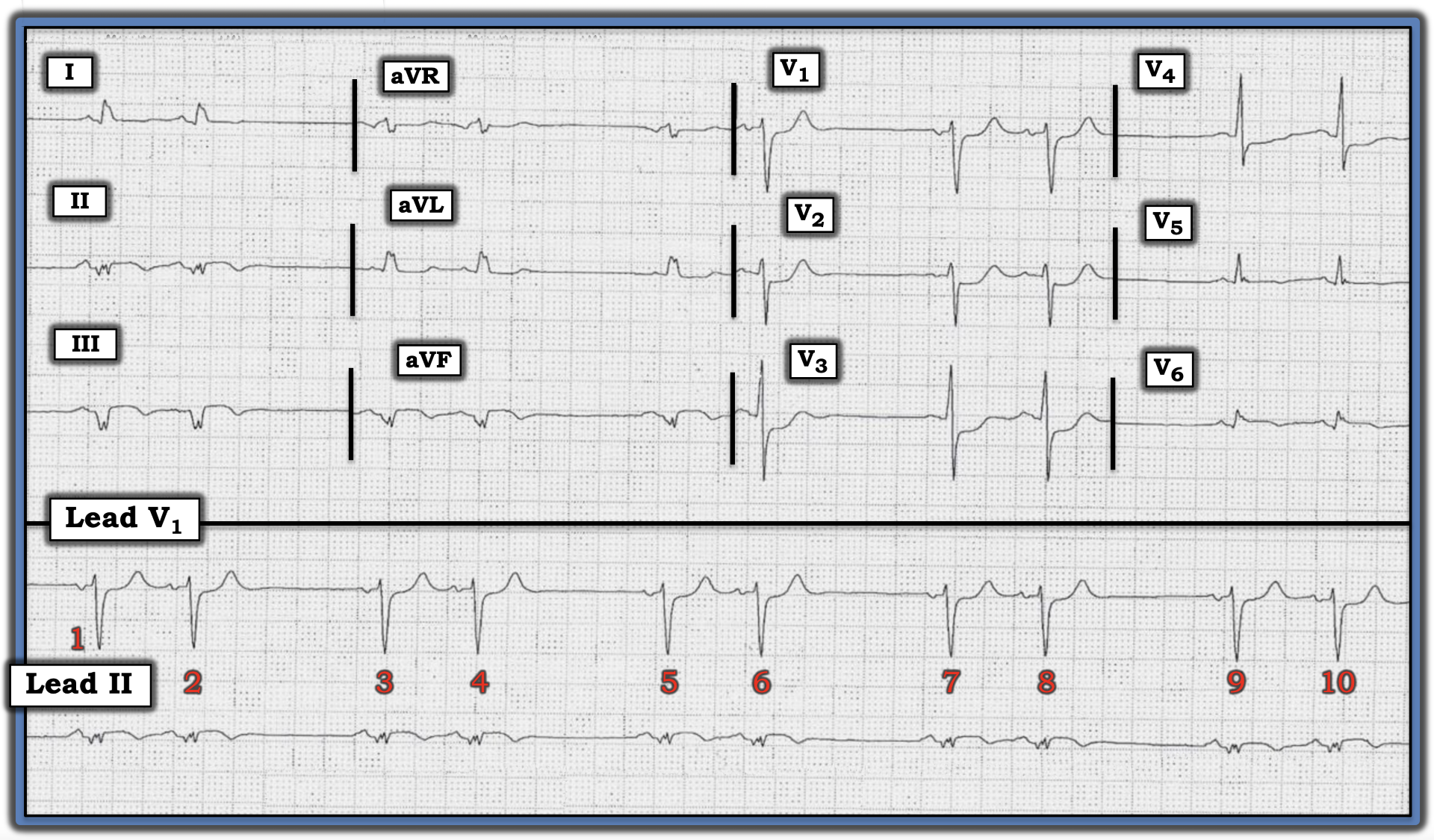
The medical providers in this case were concerned the cause of the bigeminal rhythm in the figure below was sinoatrial (SA) block. Do you agree? Are there other things to be concerned about?

Interpretation: The rhythm in this tracing fits the definition of “bigeminy” because there are repetitive groups of two beats, here with similar longer-followed-by-shorter R-R intervals.
• The underlying rhythm is sinus, as determined by the presence of an upright P wave with a constant PR interval in the long lead II rhythm strip.
• The most common cause of a bigeminal rhythm such as that shown in the figure is atrial bigeminy, in which every other beat is a premature atrial contraction (PAC).
• That said, two features in this rhythm are different than the usual case of atrial bigeminy. These two features are: i) the fact the R-R interval between the two beats in each group is precisely half the longer R-R interval between groups and ii) the similarity in P wave morphology of virtually all P waves in almost all 12 leads.
Atrial bigeminy typically results in a “reset” of the SA node, such that the longer R-R intervals usually will not be precisely twice the shorter R-R intervals. Because PACs by definition arise from an ectopic site within the atria, P wave morphology of PACs usually is readily recognized as different than the P wave morphology of sinus beats.
• In contrast, type 2 SA block with 2:1 AV conduction is characterized by a uniform P wave morphology, since all impulses arise from the SA node, and a precise doubling of longer R-R intervals compared to the shorter R-R intervals.
• That said, atrial bigeminy, in which the ectopic atrial focus arises from an atrial site close to the SA node, may manifest similar P wave morphology in most, but not all, 12 leads. Given how much more common atrial bigeminy is — and given subtle-but-real differences in P wave morphology in leads II, III, and aVF — it is most likely the rhythm in this tracing is the much more common atrial bigeminy.
NOTE: Determining the rhythm in this case is not the most important ECG finding. It turns out this patient presented with new chest pain — and the fragmented inferior lead Q waves with subtle, coved ST elevation and T wave inversion — in association with reciprocal changes in lead aVL, as well as maximal ST depression in leads V1 through V3 — suggested acute infero-postero infarction in progress.
For more information about and further discussion of this case, please visit: https://tinyurl.com/KG-Blog-339SA
