ECG Review: 1° AV Block in Lead V1
ECG Review: 1° AV Block in Lead V1
By Ken Grauer, MD
 |
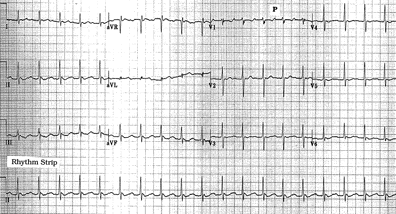
Figure. 12-lead ECG obtained from an 87-year-old man who was thought to be
in sinus tachycardia with 1° AV block.
Clinical Scenario: The 12-lead ECG shown in the Figure was obtained from an 87-year-old man, who was thought to be in sinus tachycardia with 1° AV block. Would you agree with this assessment of the rhythm?
Interpretation: The rhythm strip at the bottom of the tracing clearly shows the arrhythmia to be a regular supraventricular (narrow-complex) tachycardia at a rate of just under 120 beats/minute. It is tempting to say that the rhythm is sinus tachycardia with 1° (first degree) AV block, based on the presence of a seemingly prolonged "PR" interval in lead V1. However, this is not the correct interpretation of this rhythm.
In general, 1° AV block is uncommonly seen in the presence of sinus tachycardia. On the contrary, much of the time when a prolonged PR interval is thought to be seen with a tachycardic rhythm, a mechanism other than sinus rhythm will be the cause of this finding. This is especially true when lead II fails to show a clearly defined upright P wave. Such is the case for the Figure shown here. Thus, a second clue that the mechanism of this rhythm is unlikely to be sinus is seen in lead II, which shows no more than a poorly defined broadened and low amplitude positive deflection at the midpoint of the R-R interval. Although one cannot exclude the possibility that a sinus conducting P wave could be hidden within this low amplitude deflection, an alternative etiology for the rhythm is much more likely. Stepping back from the tracing provides the next clue—in the form of a subtle sawtooth pattern in the lead II rhythm strip (as well as in the other inferior leads). In further support of our theory that the rhythm in the Figure is atrial flutter is the ECG appearance in lead I, which shows a small rounded hump at the beginning of the ST segment, as well as an additional small rounded upright deflection preceding each QRS complex. Use of calipers allows one to walk out a consistent interval between each of these rounded deflections in lead I at a rate of 240/minute. Although the atrial rate of flutter activity in adults is most commonly closer to 300/minute, treatment with an antiarrhythmic drug may slow the atrial rate and produce flutter with 2:1 AV conduction at a ventricular rate of 120/minute as shown here.
Dr. Grauer, Professor, Assistant Director, Family Practice Residency Program, University of Florida ACLS Affiliate Faculty for Florida, is Associate Editor of Internal Medicine Alert.
Subscribe Now for Access
You have reached your article limit for the month. We hope you found our articles both enjoyable and insightful. For information on new subscriptions, product trials, alternative billing arrangements or group and site discounts please call 800-688-2421. We look forward to having you as a long-term member of the Relias Media community.
