Emergency Management of the Difficult Airway: New Techniques, Devices, and Interventional Approaches
Emergency Management of the Difficult Airway: New Techniques, Devices, and Interventional Approaches
Part II: Fiberoptic Endoscopic Techniques, Esophageal Tracheal Combitube, Gum Elastic Bougie, and Lighted Stylet
Authors: Kenneth H. Butler, DO, Associate Residency Director, University of Maryland Emergency Medicine Residency Program, University of Maryland School of Medicine, Baltimore; Brian Clyne, MD, Chief Resident, Division of Emergency Medicine, Department of Surgery, University of Maryland School of Medicine, Baltimore; and Brian D. Euerle, MD, Residency Director, University of Maryland Emergency Medicine Residency Program, University of Maryland School of Medicine, Baltimore.
Peer Reviewer: William B. Ignatoff, MD, Attending Emergency Physician; Medical Director for Adult Critical Care Transport and Transfer Referral Center, University Hospitals of Cleveland, OH; Clinical Instructor in Emergency Medicine, Case Western Reserve University, School of Medicine, Cleveland, OH.
As emphasized in Part I of this two-part series on airway management, emergency physicians must be skilled in securing an airway with techniques other than those mandated by the standard methods of oral and nasal intubation. Such alternative techniques (e.g., dual-lumen airway devices, transillumination intubation, flexible standard fiberoptics, rigid fiberoptics, and semi-rigid stylets) frequently will produce a favorable outcome in a complicated, atypical patient.
The complication rate associated with emergency intubation is significantly greater than that for routine airway management in the operating room.1 Moreover, emergency intubation is associated with cardiac arrest in more than 1% of cases.2 The most important factor in determining success or failure in airway management is the skill level of the airway manager. In this regard, the intubating physician must be familiar with various types of airway equipment and he or she must be able to select and apply the appropriate device or technique for the needs of a specific airway resuscitation.
With these critical concerns in mind, the purpose of this concluding part of our series is to present current strategies and new techniques for managing the difficult airway. Figures and tables are used to guide the physician in specific, patient-directed techniques that will improve outcomes in challenging, life-threatening emergencies.
— The EditorCombitube
The esophageal tracheal Combitube (ETC) (Kendall-Sheridan Catheter Corporation, Argyle, NY) is a double-lumen tube originally designed for blind oral intubation during cardiopulmonary resuscitation.3 The objective of this new device was to eliminate complications encountered with the esophageal obturator airway (EOA), specifically the production of airway obstruction during accidental tracheal intubation and esophageal rupture.4
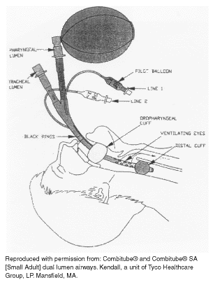
Since its inception, the ETC has been studied for in-hospital use during cardiac arrest, as a rescue device in the prehospital setting, and as a means of enabling unskilled medical personnel to provide emergency airway support.3,5-8 The device consists of two lumens, a "pharyngeal" lumen and a "tracheal" lumen, which are separated by a partition wall. One tube has an open distal end similar to an (endotracheal tube) ETT, and the other is closed at the distal end with multiple ventilating eyes proximal to its inflatable cuff. (See Figure 1.) Ventilation is possible through either lumen. Two sizes are available: 41 Fr for adult males and a 37 Fr (Combitube SA) for women and small adults.
Figure 1. The Combitube in the Esophageal Position
 |
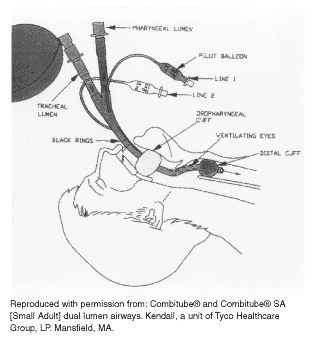
The device is inserted blindly, while the tongue and jaw are grasped between the thumb and forefinger, and positioned to a depth at which the two black ring markers are between the front teeth or alveolar ridges. The pharyngeal cuff is then inflated, sealing the tube in the posterior pharynx, limiting aspiration of oral contents, and minimizing movement. Next, the esophageal cuff is inflated, sealing the esophagus. Ventilation should then be attempted through the pharyngeal lumen, and the chest auscultated for breath sounds. If breath sounds are absent or gastric insufflation is heard (suggesting that the distal tip was inserted into the trachea), the patient should be ventilated through the tracheal lumen, and the chest again auscultated for breath sounds. (See Figure 2.) With blind insertion, the distal tip will enter the esophagus approximately 95% of the time. Tube placement can be confirmed by conventional means such as auscultation, end-tidal CO2, and self-inflating bulb.9,10
Figure 2. The Combitube in the Tracheal Position
 |
Little training time is required to use the ETC effectively. It is a useful alternative to endotracheal intubation for medical personnel untrained in laryngoscopy.6,8 In a study comparing standard endotracheal intubation by intensivists to ETC insertion by nurses, time to intubation was significantly shorter with the ETC (18.5 vs 27.2 seconds).6 Average insertion time is 10-45 seconds,11,12 and the rate of successful first-time insertion by unskilled users after brief training is 86-92%.8,13
Studies examining the adequacy of oxygenation and ventilation using the device find that the ETC provides significantly better oxygenation than an ETT and comparable PaCO2 after 20 minutes of ventilation following cardiac arrest.6,14 One report describes use of the ETC with adequate oxygenation and ventilation for up to eight hours.15
Indications for using the ETC include any clinical situation in which direct endotracheal intubation with conventional techniques is not possible. There are several potential advantages of the device: 1) It allows medical personnel without training in laryngoscopy to establish airway support in emergency situations; 2) it may obviate the need for cricothyrotomy in trauma patients with maxillofacial injuries;16 and 3) because it is inserted with the head and neck in the neutral position, the ETC may offer an advantage over endotracheal intubation (ETI) in patients with cervical spine injuries.17
The device has been used successfully in failed and difficult airway situations.18,19 The ETC probably protects against aspiration better than the laryngeal mask airway (LMA); however, this has not been studied directly. When properly positioned, the ETC allows gastric suctioning but not tracheal suctioning. Although the device was designed to be inserted blindly, insertion under direct laryngoscopy is possible and subsequent endotracheal intubation around the device can be accomplished. The ETC has a high rate of success in the prehospital setting and was preferred in one study over the LMA by emergency medical personnel.13 Each unit costs $50 and is not designed for reuse.
Contraindications include patients with intact laryngeal or pharyngeal reflexes, known esophageal disease, or corrosive ingestions. The Combitube SA, designed for small adults, should not be used in patients less than 4 feet tall. Adverse hemodynamics have been described with the ETC. Compared to the LMA and ETT, ETC insertion is associated with significant increases in heart rate, blood pressure, and plasma catecholamine levels, suggesting that the device should be avoided or used with caution in patients with cardiovascular or cerebrovascular disease.20
Despite design advances over the EOA, there still exists the potential for esophageal or pharyngeal trauma with the ETC. In one study, 18 of 40 patients (45%) had pharyngeal mucosal tears despite strict adherence to insertion guidelines. The authors postulated that the pharyngeal balloon was overinflated using manufacturer recommendations and that inflation should vary depending on the individual patient.17 There are case reports of subcutaneous emphysema due to piriform sinus perforation or esophageal laceration, apparently due to direct esophageal trauma during ETC insertion.21-23 In addition, pneumomediastinum and pneumoperitoneum have been reported after insertion.22
Despite its limitations, the literature supports claims that the ETC is an effective adjunct airway device.17,21-23 Emergency physicians should be familiar with its use and consider it in cases of unanticipated difficult intubation. Insertion of the ETC is easily learned, making it particularly appealing for use by unskilled personnel and prehospital care providers.
Lighted Stylet
First described in 1959, light-guided intubation evolved from the observation that bright light transilluminates the soft tissues of the anterior neck when placed in the trachea and does not transilluminate when placed in the esophagus.24 Using this principle, several lighted stylets or lightwand devices were developed for blind oral and nasal endotracheal intubation.
Among the newest is the Trachlight (Laerdal Medical, Armonk, NY), a lightwand device with three parts: 1) a reusable handle containing a battery pack and light source; 2) a flexible tube with a lightbulb at the distal tip; and 3) a retractable stylet within the lightwand to provide stability. (See Figure 3.) The lightwand is passed through a standard ETT and adjusted so that the lightbulb is just at the distal end of the tube. The tube is then bent to a sharp 90° angle, and insertion is performed blindly (i.e., without direct visualization of the vocal cords). The tongue and jaw are pulled forward gently from the side of the mouth, and the ETT is inserted into the back of the mouth at the midline. The tip of the ETT is then gently moved anteriorly until a bright, well-defined glow illuminates the thyroid prominence. The stylet is then retracted approximately 10 cm to allow flexibility at the tip, and the tube is advanced until the light disappears just below the sternal notch. At this point, the tip of the ETT is positioned reliably between the cords and the carina. The lightwand is then removed, followed by standard confirmation of tube placement.
Figure 3. Light-Guided Intubation with Trachlight
 |
Expertise in the technique of lighted stylet intubation is acquired quickly, and emergency physicians have used these devices with excellent success.25 One study found that in the operating room, time to intubation using a lighted stylet is significantly less compared to laryngoscopy (16 vs 20 seconds, respectively).26 Others, however, found no difference in time to intubation or success rates between lighted stylet and laryngoscopy.27,28 In a study comparing the two techniques in the operating room, all patients (50 of 50) were successfully intubated by either emergency physicians or anesthesiologists using the lighted stylet, with no significant difference in time to intubation. No major complications or airway trauma were reported.27 In the prehospital setting, 10 of 24 (48%) cases of failed intubation with direct laryngoscopy were intubated readily with the transillumination technique.29 Lighted stylets are associated with less trauma and fewer adverse hemodynamic effects when compared to conventional laryngoscopy.26,30
Indications for using the lighted stylet include patients with difficult airways due to anatomic considerations, especially patients with large overbites, restricted mouth opening, or poor dentition.31,32 The stylet can be used as a nasotracheal or orotracheal adjunct for severe facial trauma.33,34 Because insertion requires no neck or head manipulation, one special advantage may be in patients with cervical spine injury.27 In the setting of the failed airway, the lightwand can be used as intended or employed as a standard stylet to aid in direct visualization of the cords. The lighted stylet also can be used to reposition endotracheal tubes in previously intubated patients.35
There are no absolute contraindications to the use of lighted stylets in the failed or difficult airway, but limitations may be encountered in patients with known inflammatory laryngeal disorders such as epiglottitis, retropharyngeal abscess, and tracheal stenosis. They are relatively contraindicated in patients known to have laryngeal tumors, polyps, foreign bodies, or an unknown cause of upper airway compromise.35 Copious airway secretions or blood, as well as bright ambient lighting, may limit transillumination. In one study, failed lightwand intubations were attributed to a combination of factors, including thick-necked patients, operator inexperience, and bright ambient lighting.29 The degree of transillumination provided by many early lightwand devices may not be adequate for field use. In dark-skinned patients, difficulty may be encountered because of poor transillumination. Conversely, in thin, fair-skinned patients, some transillumination may be evident despite esophageal placement.25 It should be stressed that lighted stylet intubation is a blind technique, and every effort should be made to confirm tracheal placement.
The reusable handle of the Trachlight costs $200. Single-use lightwands for the device cost $20 each, and multiple-use (10 times) lightwands are available for $40 each. Overall, intubation with transillumination devices is safe and effective when used by experienced personnel during elective surgical cases. The limited data available suggest that the lighted stylet is a useful adjunct device for the failed or difficult airway in the emergency department (ED), with particular advantages in patients with facial or cervical spine trauma. As with any airway device, preparation and frequent practice are essential to maintain skills.
Fiberoptic Intubation
The use of fiberoptic endoscopy to guide intubation is a powerful tool in airway management. It has been used historically in the operating room suite by anesthesia personnel in cases of difficult airways. More recent literature describes its use in EDs by emergency physicians.36-39 A recent national survey of emergency medicine residency directors reported that 64% of the emergency medicine programs had a flexible fiberoptic bronchoscope available in their ED.40 The use of fiberoptic endoscopy in EDs may become more commonplace in the future as more emergency physicians graduate from residency programs that teach this technique.
Equipment. The fiberoptic endoscope consists of the viewing/control head, which is attached to the long thin body through which the fiberoptic bundles travel. The distal tip of the body can be moved in any direction by fingertip controls located at the head. All modern scopes contain a narrow channel running the length of the body, exiting at the tip. When the fiberoptic scope is used for intubation, this channel can be used to suction fluids away or, alternatively, to insufflate oxygen, which can blow away secretions and help oxygenate the patient.
All fiberoptic endoscopes have a light source, and there are two main types. The older, traditional type, used widely today, has a separate light source that uses AC power from a wall plug. This light source is bulky and usually kept on a small rolling cart. It is connected to the head of the endoscope by a fiberoptic cable. The advantage of this light source is that it provides a powerful, constant, reliable beam. The disadvantage is that a separate piece of equipment must be pushed around and that some physicians feel hindered by having the scope connected to the light source.
The most recently developed devices rely on a light source that contains a battery-powered unit entirely within the head of the scope. This provides the advantage of an easily transported and maneuvered scope. Potential disadvantages are less power and the need for battery replacement.
Three measurements should be considered when selecting a fiberoptic endoscope for intubations. The overall length must be sufficient such that, when the nasal route is used, the scope’s tip can be advanced sufficiently into the trachea. An overall length of 60 cm is felt to be adequate.41 The outside diameter of the body of a scope generally ranges from 3 to 5 mm. Advances in technology have resulted in smaller diameter scopes, without loss of durability, image quality, or illumination. In general, a smaller diameter scope is preferred because it is better tolerated by patients and it allows for the use of a smaller endotracheal tube. The final consideration is the diameter of the working channel. This is of relative unimportance to the intubationist, who usually will use this channel only for the insufflation of oxygen. For other uses, such as formal bronchoscopy, the channel’s diameter can be important, as it is used to pass biopsy tools and other devices.
Patient Selection. Patient selection is an important factor for fiberoptic-endoscope-guided intubation. Proper selection of patients will maximize the success rate and minimize complications. Because intubation may take several minutes to accomplish with this technique, generally it is not used with apneic patients. Fiberoptic intubation via the nasotracheal route is contraindicated in patients with severe mid-face trauma or a basilar skull fracture. Fiberoptic intubation is less likely to be successful in the patient with profuse airway bleeding and secretions. Coagulopathic patients are more likely to have complications with this technique and therefore are not good candidates.
However, fiberoptic intubation is well suited for use in the awake and breathing patient who is anticipated to have a difficult airway. The anticipated difficulty may be because of anatomic considerations, soft tissue swelling due to infection, allergic reaction, thermal injury, or a clenched jaw. In the trauma patient with a possible cervical spine injury, fiberoptic intubation can be very useful because it is reported to produce no movement of the cervical spine.36 Fiberoptic intubation may be used as the initial, primary method of controlling a patient’s airway, or it may be used as a rescue technique when the primary method has failed.
Technique. Proper preparation of the patient plays a large role in the success of the intubation. The main goals of preparation are to provide anesthesia to increase patient comfort and cooperation; to shrink mucosal membranes to permit easier passage of the endotracheal tube; and to decrease bleeding, which can make the procedure more difficult. The nasal and oral mucosa can be sprayed with 1-2% neo-synephrine or 4% lidocaine solution,36,41 or pledgets soaked in 4% lidocaine may be placed in the nose for 5 minutes.36 Alternatively, the patient can gargle with 10 mL of 4% lidocaine.36 Translaryngeal injection of 2-4 mL of 4% lidocaine solution is very effective, though few emergency physicians may be comfortable performing this procedure.39
The patient should be monitored before, during, and after the procedure, through the use of continuous pulse oximetry. Intravenous access should be obtained, and this route can be used to administer pre-procedure sedation with benzodiazepines when indicated. Finally, both the fiberoptic scope and endotracheal tube should be lubricated properly.
Fiberoptic intubation can be performed by either the nasotracheal or the orotracheal route. The nasotracheal approach will be described here because it is used far more commonly. This method is better tolerated by patients and is technically less demanding.
The adapter is removed from the end of the endotracheal tube, and the tip, followed by the body of the scope, is placed into the endotracheal tube. The endotracheal tube is slid proximally on the scope until it abuts the head. The procedure can then begin.
Procedure. The tip of the scope is introduced into the nasal passage and passed through the nasopharynx and into the oropharynx. Once these curves have been negotiated, it is normally a straight path to the vocal cords. It may be helpful to have the tongue pulled up with forceps or manually, thus elevating the epiglottis.42 Suctioning is provided as needed. The scope is then passed through the larynx until the trachea is visualized. At this point, the endotracheal tube is advanced over the scope until it is positioned properly. Care must be taken not to let the tip of the scope slip out of the trachea prematurely; otherwise, inadvertent esophageal intubation could result.37 Once the endotracheal tube is located in the trachea, the scope can be removed and ventilation started.
Location of the endotracheal tube should be confirmed by any of the standard techniques. A slight modification of this technique exists and may be preferred by some physicians. With this method, the endotracheal tube is not placed over the fiberoptic scope to start. Rather, the endotracheal tube, by itself, is first passed through the nares and advanced until the oropharynx is entered. Then the scope is passed into the endotracheal tube and advanced as outlined above. This technique is especially useful when a shorter endoscope is being used.37 After the intubation, the scope should be cleaned carefully according to the manufacturer’s instructions.
Pitfalls. Several studies have looked at the use of flexible fiberoptic guided intubations in the ED and have reported success rates of 72-87%.36-39 Failure to intubate was most often due to anatomic constraints such as narrowed nasal passages,37 to physician inexperience, and to excess blood and vomitus.36 The most commonly reported complication is bleeding.37 The median time to complete a successful fiberoptic intubation was 2 minutes in one study.39 There is an operator learning curve, as the time to intubation decreases after 9 or 10 intubations.37
Other Uses. The fiberoptic scope has uses in the ED other than its use in intubations. A variety of airway problems can be evaluated. In the patient with blunt laryngeal trauma, for example, the fiberoptic scope can be used to examine the vocal cords, supraglottic structures, and subglottic area.36 The same examination can be done in the burned patient or the patient with a foreign body ingestion.
In summary, the use of flexible fiberoptic-guided intubation can be an important resource for the emergency physician. Judicious patient selection can help increase the likelihood of success. Proper preparation of the patient and the equipment also can help ensure success as well as decrease complications. The emergency physician may employ fiberoptic intubation both as a primary method of intubation in the patient with a difficult airway and a rescue technique when other methods have failed.
The WuScope
The WuScope (Achi Corporation, Fremont, CA) is a new fiberoptic intubating device that combines a rigid laryngoscope blade with a flexible fiberscope. Introduced in 1993 by Wu and Chou, it is designed to facilitate endotracheal intubation with the patient’s head in the neutral position.42 The rigid blade has three components: a handle, a main blade, and a bivalve element. (See Figure 4.)
Figure 4. The Combination Intubating Device Developed by Wu
 |
The handle is cone-shaped and accepts the fiberscope body. The main blade and bivalve element connect to form a tubular exoskeleton with two passageways, one for the ETT and one for the fiberscope. (See Figure 5.)
Figure 5. Rear View of the Three Parts of the Rigid Blade Portion of the Device
 |
It is recommended that a suction catheter be placed through the ETT as a guide during intubation.
With the endotracheal tube and fiberscope secured, the device is held in the intubator’s left hand and inserted into the patient’s mouth at the midline. While the posterior pharynx is visualized through the eyepiece of the fiberscope (or on color video monitor), the handle is rotated toward the operator and the blade advanced toward the larynx. (See Figure 6.)
Figure 6. The Device as Used in the Orotracheal Intubation
 |
The suction catheter, acting as a guide, is advanced into the glottis. The distal main blade is adjusted to align the ETT with the glottis, and the ETT is then advanced over the suction catheter into the trachea. The bivalve element is removed first from the patient’s mouth, followed together by the handle, main blade, and fiberscope as a unit. Nasotracheal intubation is performed by inserting the device into the mouth without the bivalve element attached.
Several design features make the WuScope unique and offer potential advantages over other laryngoscopes, rigid or flexible. The modified handle-to-blade angle of 110°, compared with the 90° conventional laryngoscopes, may provide an advantage during blade insertion in obese, short-necked, or barrel-chested patients. The blade contour is similar to that of an oral airway and allows insertion into the larynx with minimal head extension or tongue lifting, making it suitable for the awake or cervical spine-injured patient. An oxygen port and channel on the main blade allow supplemental oxygenation during intubation, and suctioning is possible through the catheter that guides the ETT. The tubular blade structure protects the fiberscope from secretions, blood, and redundant soft tissue.
Research indicates that the WuScope is an effective device to facilitate orotracheal or nasotracheal intubation in routine or difficult airway situations. Smith found that use of the WuScope was associated with relatively easy glottic exposure and tracheal intubation in patients with difficult airways.43 Wu reports successful use in 48 patients with Mallampati classification III and IV, including 11 patients with adverse anatomic features and previously documented difficult airways. It also has been used as a rescue device by anesthesiologists in cases of failed endotracheal intubation.42 In patients with cervical spine stabilization, laryngeal view with the WuScope is superior to conventional laryngoscopy with significantly less head extension.44,45
Contraindications to the WuScope include patients with limited mouth opening and extreme and fixed airway deformities. If patients cannot open at least 20 mm to accommodate the device or have upper airway deformities precluding insertion, an alternate method should be chosen. Technical difficulties observed with the WuScope include fogging of the fiberscope and poor visualization due to copious secretions.45 The potential for oropharyngeal trauma is similar to that with other rigid laryngoscopes. Perhaps the most significant limitation is the high initial cost of the device. The cost of the basic package, which includes the fiberscope, blades, and handle, is $9,800. Although the WuScope is reusable, the maintenance, repair, and training costs may make the device prohibitively expensive for many EDs. The need for a fiberoptic light source makes it impractical for prehospital use; however, a modified battery-powered source will be available soon.
Gum Elastic Bougie
The gum elastic bougie (also known as a tracheal tube introducer or Eschmann stylet) is a long, semirigid malleable device made of woven polyester with a resin coating. This device was described by Macintosh in 1949 as an aid to intubation.46 Simple in its design, the gum elastic bougie is useful for the unanticipated difficult airway and for patients with suspected cervical spine injury. The standard size device is 60 cm long and 15 Fr in diameter, with a 40° angle 3.5 cm from its distal tip.47
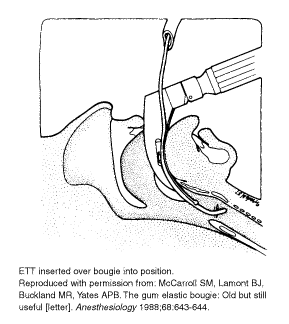
During cases of absent or partial visualization of the vocal cords, the gum elastic bougie is lubricated and passed posterior to the epiglottis with the distal tip angled anteriorly. (See Figure 7.)
Figure 7. Bougie Directed into the Trachea
 |
Reliable signs of tracheal placement are clicks felt as the tip of the bougie passes over the tracheal cartilage rings.48 With esophageal placement, clicks are not felt and the device can be advanced without hold up for more than 45 cm. With the bougie stabilized in the trachea and the laryngoscope still held in the mouth, an ETT is threaded over the bougie into the trachea. Passage of the ETT is made easier by rotating the tube 90° counterclockwise, keeping the bevel of the tube posterior.49 Some suggest the bougie is best used in conjunction with an anterior commissure laryngoscope.50
Although few data exist regarding bougie use in the ED, there are reports of successful use in patients with airway edema, neck trauma, or cervical spine immobilization.51-53 It has been used as an adjunct to cricothyrotomy in the prehospital setting.54 A modification of the device is the jet stylet introducer, a hollowed-out bougie with capacity for continuous end-tidal CO2 monitoring. This device reliably distinguishes tracheal from esophageal placement before passing an ETT.55
Advantages of the bougie are its simplicity, expense ($75 each), and reusability. The tip of the device is malleable and can be curved as needed. Local trauma to the airway is a risk but can be avoided with gentle technique. An inexpensive, easily used device for the unanticipated difficult airway, the gum elastic bougie should be kept at arm’s length during every intubation.
Summary
Experienced clinicians understand that any intubation may be difficult and may result in poor clinical outcomes. Recognition of the predictors of a difficult airway, proper preparation, and the ability to use alternative devices may be the difference between life and death.
Emergency physicians should always provide the ABCs in patient stabilization, and airway management is the mainstay of emergency intervention. If we are to continue to hold ourselves responsible for emergency care, we should possess the knowledge and equipment to manage patients with complicated airways. As technology and clinical data continue to advance, and with the development of devices for airway management, traditional laryngoscopic intubation may not remain the standard of care for every airway.
References
1. Mascia MF, Mattasko MT. Emergency airway management by anesthesiologists. Anesthesiology 1993;79:A1054.
2. Mort TC. Incidence and risks leading to cardiac arrest following emergency intubation. Crit Care Med 1994;22:A137.
3. Frass M, Frenzer R, Zdrahal F, et al. The esophageal tracheal combitube: Preliminary results with a new airway for CPR. Ann Emerg Med 1987;16:768-772.
4. Pepe PE, Zachariah BS, Chandra NC. Invasive airway techniques in resuscitation. Ann Emerg Med 1993;22:393-403.
5. Frass M, Frenzer R, Rauscha F, et al. Ventilation with the esophageal tracheal combitube in cardiopulmonary resuscitation: Promptness and effectiveness. Chest 1988;93:781-784.
6. Staudinger T, Brugger S, Watschinger B, et al. Emergency intubation with the combitube: Comparison with the endotracheal airway. Ann Emerg Med 1993;22:1573-1575.
7. Atherton G, Johnson J. Ability of paramedics to use the combitube in prehospital cardiac arrest. Ann Emerg Med 1993;22:1263-1268.
8. Yardy N, Hancox D, Strang T. A comparison of two airway aids for emergency use by unskilled personnel: The combitube and laryngeal mask. Anaesthesia 1999;54:181-183.
9. Butler BD, Little T, Drtil S, et al. Combined use of the esophageal tracheal combitube with a colormetric carbon dioxide detector for emergency intubation/ventilation. J Clin Monit 1995;11:311.
10. Salem MR, Wafai Y, Baraka A, et al. Effectiveness of the self-inflating bulb for verification of proper placement of the Esophageal Tracheal Combitube. Anesth Analg 1995;80:122-126.
11. Frass M, Reinhard F, Rauscha F, et al. Evaluation of esophageal tracheal combitube in cardiopulmonary resuscitation. Crit Care Med 1986;15:609-611.
12. Calkins MD, Robinson T. Combat trauma airway management: Endotracheal intubation versus laryngeal mask airway versus combitube use by Navy SEAL and Reconnaissance combat corpsmen. J Trauma 1999;46:927-932.
13. Rumball CJ, MacDonald D. The PTL, Combitube, laryngeal mask, and oral airway: A randomized prehospital comparative study of ventilatory device effectiveness and cost-effectiveness in 470 cases of cardiorespiratory arrest. Prehospital Emergency Care 1997;1:58-59.
14. Frass M, Rodler S, Frenzer R, et al. Esophageal Tracheal Combitube, endotracheal airway, and mask: Comparison of ventilatory pressure curves. J Trauma 1989;29:1476-1479.
15. Frass M, Frenzer R, Mayer G, et al. Mechanical ventilation with the esophageal tracheal combitube (ETC) in the intensive care unit. Arch Emerg Med 1987;4:219-225.
16. Blostein P, Koestner A, Hoak S. Failed rapid sequence intubation in trauma patients: Esophageal Tracheal Combitube is a useful adjunct. J Trauma 1998;44:534-537.
17. Mercer MH, Gabbott DA. The influence of neck position on ventilation using the Combitube airway. Anaesthesia 1998;53:146-150.
18. Bigenzahn W, Pesau B, Frass M. Emergency ventilation using the Combitube in cases of difficult intubation. Eur Arch Otorhynolaryngol 1991;248:129.
19. Klauser R, Roggla G, Pidlich J, et al. Massive upper airway bleeding after thrombolytic therapy: Successful airway management with the Combitube. Ann Emerg Med 1992;21:431-433.
20. Oczenski W, Krenn H, Dahaba A, et al. Hemodynamic and catecholamine stress responses to insertion of the Combitube, laryngeal mask airway or tracheal intubation. Anesth Analg 1999;88:1389-1394.
21. Richards CF. Piriform sinus perforation during esophageal-tracheal Combitube placement. J Emerg Med 1998;16:37-39.
22. Vezina D, Lessard MR, Bussieres J, et al. Complications associated with the use of the esophageal-tracheal Combitube. Can J Anesth 1998;45:823-824.
23. Klein H, Williamson M, Sue-Ling HM, et al. Esophageal rupture associated with the use of the Combitube. Anesth Analg 1997;85:937-939.
24. Yamamura H, Yamamoto T, Kamiyama M. Device for blind nasal intubation. Anesthesiology 1959;20:221.
25. Ainsworth QP, Howells TH. Transilluminated tracheal intubation. Br J Anaesth 1989;62:494-497.
26. Hung OR, Pytka S, Morris I, et al. Clinical trial of a new lightwand device (Trachlight) to intubate the trachea. Anesthesiology 1995;83:509-514.
27. Ellis DG, Jakymec A, Kaplan RM, et al. Guided orotracheal intubation in the operating room using a lighted stylet: A comparison with direct laryngoscopic technique. Anesthesiology 1986;64:823-826.
28. Ellis DG, Stewart RD, Kaplan RM, et al. Success rates of blind orotracheal intubation using a transillumination technique with a lighted stylet. Ann Emerg Med 1986;15:138-142.
29. Vollmer TP, Stewart RD, Paris PM, et al. Use of a lighted stylet for guided orotracheal intubation in the prehospital setting. Ann Emerg Med 1985;14:324-328.
30. Knight RG, Castro T, Rastrelli AJ, et al. Arterial blood pressure and heart rate response to lighted stylet or direct laryngoscopy for endotracheal intubation. Anesthesiology 1988;69:269-272.
31. Hung OR, Pytka S, Morris I, et al. Lightwand intubation: II. Clinical trial of a new lightwand for tracheal intubation in patients with difficult airways. Can J Anaesth 1995;42:826-830.
32. Crosby ET, Cooper RM, Douglas MJ, et al. The unanticipated difficult airway with recommendations for management. Can J Anesth 1998;45:757-776.
33. Verdile VP, Heller MB, Paris PM, et al. Nasotracheal intubation in traumatic craniofacial dislocation: Use of the lighted stylet. Am J Emerg Med 1988;6:39-41.
34. Verdile VP, Chiang JL, Bedger R, et al. Nasotracheal intubation using a flexible lighted stylet. Ann Emerg Med 1990;19:506-510.
35. Hung OR, Stewart RD. Lightwand intubation: I. A new lightwand device. Can J Anaesth 1995;42:820-825.
36. Schafermeyer RW. Fiberoptic laryngoscopy in the emergency department. Am J Emerg Med 1984;2:160-163.
37. Delaney KA, Hessler R. Emergency flexible fiberoptic nasotracheal intubation: A report of 60 cases. Ann Emerg Med 1988;17:919-926.
38. Mlinek EJ Jr, Clinton JE, Plummer D, et al. Fiberoptic intubation in the emergency department. Ann Emerg Med 1990;19:359-362.
39. Afilalo M, Guttman A, Stern E, et al. Fiberoptic intubation in the emergency department: A case series. J Emerg Med 1993;11:387-391.
40. Levitan RM, Kush S, Hollander JE. Devices for difficult airway management in academic emergency departments: Results of a national survey. Ann Emerg Med 1999;33:694-698.
41. Edens ET, Sia RL. Flexible fiberoptic endoscopy in difficulty intubations. Ann Otol Rhinol Laryngol 1981;90:307-309.
42. Smith CE, Sidhu TS, Lever J, et al. The complexity of tracheal intubation using rigid fiberoptic laryngoscopy (WuScope). Anesth Analg 1999;89:236-239.
43. Wu TL, Chou HC. A new laryngoscope: The combination intubating device. Anesthesiology 1994;81:1085-1087.
44. Sandhu NS, Schaffer S, Capan LM, et al. Comparison of the WuScope and Macintosh #3 blade in normal and cervical spine stabilized patients. Anesthesiology 1999;91:A480.
45. Smith CE, Pinchak AB, Sidhu TS, et al. Evaluation of tracheal intubation difficulty in patients with cervical spine immobilization. Anesthesiology 1999;91:1253-1259.
46. Macintosh RR. An aid to oral intubation. Br Med J 1949;1:28.
47. McCarroll SM, Lamont BJ, Buckland MR, et al. The gum-elastic bougie: Old but still useful [letter]. Anesthesiology 1988;68:643-644.
48. Kidd JF, Dyson A, Latto IP. Successful difficult intubation: Use of the gum elastic bougie. Anaesthesia 1988;43:437-438.
49. Dogra S, Falconer R, Latto IP. Successful difficult intubation: Tracheal tube placement over a gum-elastic bougie. Anaesthesia 1990;45:774-776.
50. Sofferman RA, Johnson DL, Spencer RF. Lost airway during anesthesia induction: Alternatives for management. Laryngoscope 1997;107:1476-1482.
51. Nolan JP, Wilson ME. Orotracheal intubation in patients with potential cervical spine injuries: An indication for the gum elastic bougie. Anaesthesia 1993;48:630-633.
52. Randalls B, Toomey PJ. Laryngeal oedema from a neck haematoma. Anaesthesia 1990;45:850-852.
53. Groves J, Edwards N, Hood G. Difficult intubation following thoracic trauma. Anaesthesia 1994;49:698-699.
54. Morris A, Lockey D, Coats T. Fat necks: Modification of a standard airway protocol in the prehospital environment. Resuscitation 1997;35:253-254.
55. Spencer RF, Rathmell JP, Viscomi CM. A new method for difficult endotracheal intubation: The use of a jet stylet introducer and capnography. Anesth Analg 1995;81:1079-1083.
Subscribe Now for Access
You have reached your article limit for the month. We hope you found our articles both enjoyable and insightful. For information on new subscriptions, product trials, alternative billing arrangements or group and site discounts please call 800-688-2421. We look forward to having you as a long-term member of the Relias Media community.
