Emergency Ultrasound, Part II: Diagnosis of Deep Venous Thrombosis
Part II: Diagnosis of Deep Venous Thrombosis
Authors: Christopher L. Moore, MD, RDMS, Assistant Professor, Section of Emergency Medicine, Yale University School of Medicine, New Haven, CT; Michael J. Lambert, MD, RDMS, Clinical Assistant Professor, Department of Emergency Medicine, University of Illinois College of Medicine; Fellowship Director, Emergency Ultrasound, Resurrection Medical Center, Chicago, IL.
Peer Reviewers: Anthony Macasaet, MD, Assistant Professor and Vice Chair, Director Emergency Medicine Ultrasound, Department of Emergency Medicine, Chicago Medical School, Chicago, IL; Kenneth H. Butler, DO, Associate Residency Director, University of Maryland Emergency Medicine Residency Program, University of Maryland School of Medicine, Baltimore.
Deep vein thrombosis (DVT) is a common and potentially life-threatening peripheral vascular disease that emergency physicians (EPs) frequently consider, diagnose, and treat in their practice. The typical patient at risk for DVT presents with unilateral lower extremity swelling, pain, or discoloration. The definitive diagnosis often is impossible to ascertain based upon clinical findings alone, as numerous nonthrombotic conditions share similar signs and symptoms. Unless an alternative diagnosis confidently can be established with a high degree of certainty, EPs typically rely upon ultrasound (US) imaging to rule out DVT.
Frustrated with the time delays and availability of ultrasound imaging after-hours, more and more EPs are acquiring the skills to perform and interpret lower extremity US. Although evaluation for DVT is not considered a primary application in emergency medicine US, many EPs already have recognized the benefit of adding this particular application to their arsenal.
The purpose of this article is to review the anatomy and pathophysiology of DVT as it relates to ultrasonographic imaging, both to provide a deeper understanding of the disease and to serve as an introduction to the performance of DVT ultrasound by the EP.—The Editor
Incidence
The exact incidence of DVT is unknown. Existing data suggest that about 80 cases per 100,000 persons occur annually. Approximately 1 person in 20 develops DVT during her or his lifetime, and 600,000 hospitalizations for DVT occur annually in the United States.1 Although frequently subclinical, DVT invariably precedes the development of pulmonary embolism, estimated to occur in more than 500,000 patients per year in the United States, 11% of whom die within the first hour.
Diagnosis
The diagnosis of DVT has evolved significantly since its first documentation. While venography still is widely considered the gold standard, this invasive and time-consuming test is not technically adequate in a significant percentage of patients1 and now is widely reserved for patients in whom doubt exists following other diagnostic studies. For the symptomatic emergency department (ED) patient, US is the initial study of choice.2
Pooled results have shown US performed by radiologists to be 95% sensitive and 98% specific in the diagnosis of lower extremity DVT in symptomatic patients.2,3 The sensitivity is poorer (around 59%) when US is used for screening patients who are at high risk (post-op, etc.) but asymptomatic.4
Anatomy
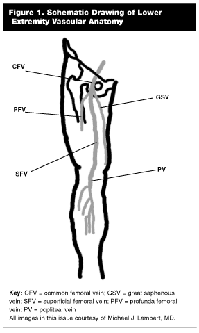
To understand the use of US in the diagnosis of DVT, it is essential to understand the anatomy and terminology of the deep venous system. While there also are deep veins in the upper extremities, this review will cover the lower extremity anatomy. (See Figure 1.)

From proximal to distal, as the iliac vein crosses below the inguinal ligament it becomes the common femoral vein. This vein is medial to the artery in the proximal thigh. The greater saphenous vein, which is a superficial vein on the medial side of the leg, usually joins the common femoral vein in the upper thigh. As the common femoral vein continues distally, it goes deeper and moves somewhat laterally relative to the artery to a position almost directly below the artery, where it bifurcates into the deep (profunda) femoral vein and the superficial femoral vein. Only the very proximal portions of the deep (profunda) femoral vein may be available to US interrogation, if at all.
The superficial femoral vein continues through the medial thigh to the adductor canal. It is important to note that the superficial femoral vein, while more superficial than the deep femoral vein, is a misnomer in that it is both part of the deep venous system and relatively deep compared to such veins as the saphenous. As the superficial femoral vein passes through the adductor canal, it becomes the popliteal vein. At this point, in the popliteal fossa, the vein is anatomically posterior, though it is closer to the skin and, thus, appears more superficial when imaged by an US probe placed in the popliteal fossa. The lesser saphenous, a superficial vein, inserts in the popliteal vein in the mid- to proximal portion.
As the popliteal vein continues distally, the anterior tibial vein is the first branch, which then runs in the anterior tibial compartment adjacent to the interosseous membrane. Following take-off of the anterior tibial vein, the popliteal vein bifurcates into the peroneal and posterior tibial veins. The anterior tibial, posterior tibial, and peroneal veins are considered deep calf veins.
Ultrasound Interrogation
The array of options included for US evaluation of the lower extremities includes: B-mode visualization of clot formation and compressibility, color flow and duplex imaging, and indirect measures such as response to respiration, valsalva, and augmentation maneuvers.
A complete ultrasound of the deep venous system includes evaluation of the common femoral, superficial femoral, popliteal, and calf veins.5 In practice, the evaluation of calf veins distal to the bifurcation of the popliteal vein frequently is omitted.2,6,7 Calf veins may be imaged with the aid of color flow Doppler, but sensitivity is variable and detection requires a significant increase in the time for performance of the study.2,4,8 While treatment of isolated calf vein thrombosis is controversial, the poor sensitivity of US for calf vein thrombosis should be noted, as up to 20% of calf vein thromboses may propagate proximally.9 This is the basis for the recommendation that a repeat US examination be performed at one week in patients with an initially negative examination who are judged to be at intermediate or high risk or who remain symptomatic.3,7,10 However, isolated calf vein thrombosis has not been shown to cause fatal pulmonary embolism, and propagation invariably occurs before embolism.9
The most reliable component of the US examination for DVT has been found to be compression ultrasonography. (See Figure 2.)

Using a linear probe in the 5-7.5 MHz frequency range, the veins are interrogated from the anterior surface of the thigh along their length at 2-3 cm intervals in the transverse (and occasionally longitudinal) planes. (See Figure 3.)

Gentle compression (see Figure 4) along the vein should cause the clot-free vein to collapse completely.

The corresponding B-mode (also known as 2-D or gray-scale) image is displayed on the ultrasound monitor screen. (See Figures 5 and 6.)


Absence of compression (vein walls fail to contact with adequate compression over vessel) indirectly indicates the presence of a clot in the lumen. (See Figure 7.)

Occasionally, a clot may be visualized directly as a slightly hyperechoic image in the lumen, a specific but not sensitive sign of a clot. (See Figure 8.)

Typically, the thigh is evaluated with the patient supine, and the popliteal vessels with either the knee bent slightly or the patient in a decubitus or prone position. (See Figure 9.)

The literature has demonstrated that it is reasonably safe to withhold anticoagulation therapy from patients with a negative compression US. In a study of 1022 patients with normal compression US (common femoral to popliteal veins evaluated) followed for more than eight months, three patients returned with DVT and two died from pulmonary embolism.7
Whether the use of compression US of the lower extremities can be abbreviated to include only the common femoral and popliteal veins is controversial. This is considered a "limited compression ultrasound," and thus does not include the superficial femoral vein. Post-mortem and venography studies have shown that DVT appears invariably to involve the common femoral vein or the popliteal vein, and does not occur as an isolated superficial femoral vein clot.11 With this in mind, one study showed an accuracy of 100% and a decrease in the time to perform the US by 9.7 minutes or 54% when using the abbreviated method.12 However, a larger, prospective US study of 755 patients reported six clots isolated to the superficial femoral veins (4.6% of all patients with DVT, 0.8% of all patients studied) and concluded that abbreviating the examination would sacrifice diagnostic accuracy unacceptably.13 However, this study included all patients referred for US for any reason (i.e., asymptomatic after surgery). The incidence and prognosis of isolated common femoral vein thrombosis in a population of symptomatic ED patients is not known.
The addition of Doppler frequently is used to enhance visualization and diagnosis of a deep venous clot. Doppler is a term that encompasses the use of US in the evaluation of a moving target, in this case blood in the venous system, and includes color flow and spectral or "duplex" Doppler.
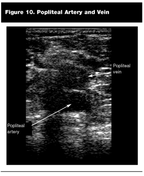
Color flow Doppler is the representation of flow toward or away from the transducer using red and blue. A normal clot-free vein will fill with color throughout the lumen in a phasic manner. The use of color flow may help to localize difficult-to-find vessels in uncooperative or obese patients, and may detect a clot in areas that are difficult to compress, such as at the inguinal ligament or where the superficial femoral vein passes through the adductor canal. Presence of a clot is identified when a luminal filling defect occurs. (See Figure 10.) While the use of color flow typically is a complementary technique to compression ultrasonography, it has been validated as an independent modality.14

Spectral Doppler is a modality that can quantify flow velocity and typically is represented with time on the x-axis and velocity (positive or negative) on the y-axis. (See Figure 11.)

It is called duplex because both the B-mode image and the spectral Doppler waveform are displayed simultaneously. Use of pulsed wave Doppler allows selection of a sample volume (gated volume) on the B-mode image that then is represented on a separate part of the screen. In patients who are free of clot, spontaneous flow variation should be evident at rest and should show phasic variation with respiration, valsalva, or squeezing of the distal calf veins (augmentation). Valsalva also should cause an increase of 50% or greater in the size of the common femoral vein on B-mode imaging. Absence of these with normal direct compressibility is evidence of a clot either proximal or distal to the area being evaluated. (See Figures 12 and 13.) While the use of duplex may enhance the US evaluation, it rarely is used alone to diagnose DVT without other direct signs by compression or color-flow examination.2


Ultrasound by EPs for DVT
The majority of the literature, and that discussed above, is based on the evaluation for DVT in a vascular laboratory with a full array of machines, trained sonographers, and physicians skilled in US interpretation. In an ideal world, all of this would be available to the EP on an immediate and inexpensive basis. While many hospitals maintain such resources on an on-call basis 24 hours per day, seven days per week, in even the best situation evaluation for DVT in off-hours is time-consuming and resource intensive, if it is available at all.15 This raises the question of the performance of US for DVT by EPs.
The literature for ultrasonographic detection of DVT by EPs is not extensive. A 1990 study described the use of a handheld Doppler stethoscope by EPs and described a sensitivity of 85% with a specificity of 79% compared with venography, although there were a high number of equivocal studies that were not included in the analysis.16 This technique is highly operator-dependent and retains only historical significance.2
Another study investigated the training and performance of lower extremity US by two EPs during off-hours.17 Following training with the radiology department, these physicians studied 15 patients and found a 100% sensitivity and a 75% specificity based on follow up radiology results. Time to complete the examination was not noted.
A study in 2000 consisted of 112 patients who were evaluated in the ED for suspected DVT. EPs performed compression ultrasonography of the femoral and popliteal veins, with assistance from color flow Doppler and augmentation maneuvers (i.e., squeezing the calf). Thirty-four patients were found to have DVT, and results were found to have excellent agreement with vascular laboratory US studies completed within eight hours (kappa of 0.9, 98% agreement). Median time for EPs to complete the study was 3 minutes 28 seconds. Limitations included lack of a true gold standard and limited long term follow-up for outcome based on EP diagnosis.15
Studies such as this suggest that the EP may be able to utilize US quickly and effectively for the majority of patients presenting with symptoms of DVT. In even the best circumstances, US is not a perfect test for DVT. It is reasonable to suppose that the judicious use of US by EPs in addition to close follow-up could be helpful in the evaluation of DVT in the ED.
Recently, studies have suggested that D-dimer may be used in the assessment of DVT with a high degree of negative predictive value,6 and is especially good when paired with low pre-test probability.10,18 In addition, the use of pretest probability paired with a negative US may reliably exclude DVT.3 It is possible that the performance of compression US for DVT by the EP in conjunction with clinical suspicion or a test such as D-dimer could be used to safely withhold anticoagulant therapy, although this awaits prospective validation.
In addition to the evaluation of DVT per se, US of the lower extremities may play a role in the emergent evaluation of patients presenting with suspected pulmonary embolism (PE), especially those in extremis. While as many as 29% of patients with documented PE have no DVT on venography,2 the presence of DVT by ultrasound provides evidence that a symptomatic patient has suffered a PE, and provides validation for the use of heparin or even thrombolytics in the unstable patient with a high clinical suspicion. Frazee and Snoey have recommended the combination of emergent transthoracic echocardiography with DVT for the patient suspected to have suffered a massive PE.1
It also should be noted that US, either done in radiology or by the EP, may reveal an alternate diagnosis when DVT is suspected. Popliteal or Baker’s cysts (See Figure 15) appear as smooth, walled, echo-free masses located medially in the popliteal fossa, occasionally with septations.19

Popliteal artery aneurysms (See Figure 16) occur when the vessel exceeds a diameter of 1.1 cm, and appear as an echo-free mass located centrally in the popliteal fossa continuous with the popliteal artery.

Doppler may establish flow in the aneurysm (See Figure 17), although presence of clot (more echogenic) may eliminate flow.19 Other diagnoses that may be made by US include abscesses, hematomas, tumors, lymphadenopathy, and muscular or tendinous inflammation.20

Conclusion
US, and specifically compression ultrasonography, is the diagnostic test of choice for evaluation of the ED patient with suspected DVT. As with any skill, there is a learning curve, and the performance of US for DVT by EPs should be approached judiciously. Knowledge of the technique, however, may provide a tool that is otherwise unavailable in a timely manner. In any case, knowledge of the performance and limitations of lower extremity US will improve diagnosis and treatment of this important disease.
References
1. Frazee BW, Snoey ER. Diagnostic role of ED ultrasound in deep venous thrombosis and pulmonary embolism. Am J Emerg Med 1999;17:271-278.
2. Cronan JJ. Venous thromboembolic disease: The role of US. Radiology 1993;186:619-630.
3. Wells PS, Anderson DR, Bormanis J, et al. Value of assessment of pretest probability of deep-vein thrombosis in clinical management. Lancet 1997;350:1795-1798.
4. Lewis BD. The peripheral veins. In: Rumack CM, Wilson SR, Charboneau JW, eds. Diagnostic Ultrasound. St. Louis, MO; Mosby 1998: 943-958.
5. AIUM Standards for performance of the vascular/Doppler ultrasound examination. 1992. http://www.aium.org/consumer/standards/vascular.asp.
6. Birdwell B. Recent clinical trials in the diagnosis of deep-vein thrombosis. Curr Opin Hematol 1999;6:275-279.
7. Vaccaro JP, Cronan JJ, Dorfman GS. Outcome analysis of patients with normal compression US examinations. Radiology 1990;175: 645-649.
8. Rose SC, Zwiebel WJ, Nelson BD, et al. Symptomatic lower extremity deep venous thrombosis: Accuracy, limitations, and role of color duplex flow imaging in diagnosis. Radiology 1990; 175: 639-644.
9. Philbrick JT, Becker DM. Calf deep venous thrombosis. Arch Int Med 1998;148:2131-2138.
10. Lensing AWA, Prandoni P, Prins MH, et al. Deep-vein thrombosis. Lancet 1999;353:479-485.
11. Cogo A, Lensing A, Prandoni P, et al. Distribution of thrombosis in patients with symptomatic deep vein thrombosis. Arch Int Med 1993;153:2777-2780.
12. Pezzullo JA, Perkins AB, Cronan JJ. Symptomatic deep vein thrombosis: diagnosis with limited compression US. Radiology 1996; 198:67-70.
13. Frederick MG, Hertzber BS, Kliewer MA, et al. Can the US examination for lower extremity deep venous thrombosis be abbreviated? A prospective study of 755 examinations. Radiology 1996;199: 45-47.
14. Lewis BD, James EM, Welch TJ, et al. Diagnosis of acute deep venous thrombosis of the lower extremities: Prospective evaluation of color Doppler flow imaging versus venography. Radiology 1994; 192:651-655.
15. Blaivas M, Lambert MJ, Harwood RA, et al. Lower-extremity Doppler for deep venous thrombosis—can emergency physicians be accurate and fast? Acad Emerg Med 2000;7:120-126.
16. Turnbull TL, Dymowski JJ, Zalut TE. Prospective study of hand-held Doppler ultrasonography by emergency physicians in the evaluation of suspected deep vein thrombosis. Ann Emerg Med 1990; 19:691-695.
17. Jolly BT, Massarin E, Pigman EC. Color Doppler ultrasonography by emergency physicians for the diagnosis of acute deep venous thrombosis. Acad Emerg Med 1997; 4:129-132.
18. Anderson DR, Wells PS, Stiell I, et al. Management of patients with suspected deep vein thrombosis in the emergency department: Combining use of a clinical diagnosis model with D-dimer testing. J Emerg Med 2000;19:225-230.
19. Pathria MN, Zlatkin M, Sartoris DJ, et al. Ultrasonography of the popliteal fossa and lower extremities. Radiol Clin N Am 1988; 26:77-85.
20. Pini M, Marchini L, Giordano A. Diagnostic strategies in venous thromboembolism. Haematologica 1999;84:535-540.
Although evaluation for DVT is not considered a primary application in emergency medicine ultrasound, many emergency physicians already have recognized the benefit of adding this particular application to their arsenal.
Subscribe Now for Access
You have reached your article limit for the month. We hope you found our articles both enjoyable and insightful. For information on new subscriptions, product trials, alternative billing arrangements or group and site discounts please call 800-688-2421. We look forward to having you as a long-term member of the Relias Media community.
