Pediatric Major Trauma
Module 3: Pediatric Trauma
Author
Aaron Leetch, MD, Assistant Professor, Departments of Emergency Medicine and Pediatrics; Associate Program Director, Emergency Medicine/Pediatrics Residency, University of Arizona, Tucson, AZ
Dr. Leetch reports no financial relationships relevant to this field of study.
Core Content Outline: Major Trauma
General principles
- Recognize common patterns of injury in children with major trauma with respect to anatomic and physiologic differences by age
- Recognize response to injury in children with major trauma with respect to anatomic and physiologic differences by age
- Know the importance of mechanisms of injury in the evaluation of children with major trauma
- Understand priorities in the management of children with major trauma
- Know triage principles in the management of victims of major trauma
- Understand the importance of thermal regulation in the management of children with major trauma
- Understand the principles of primary versus secondary survey
- Understand the importance of appropriate fluid resuscitation in the management of children with major trauma
- Understand the importance of appropriate airway management in children with major trauma
Evaluation and stabilization
Primary survey
- Recognize airway obstruction in children with major trauma
- Anticipate the risk of cervical spine injury associated with major trauma
- Understand the concept that cervical cord injury can occur in the absence of a radiologic abnormality
- Know causes of acute cardiopulmonary collapse after major trauma
- Distinguish causes of shock in the trauma patient
- Know methods of rapid assessment of the central nervous system
- Know causes of delayed acute cardiopulmonary collapse after major trauma
Trauma resuscitation
- Plan the management of a child with an obstructed airway in the setting of major trauma
- Know the components of rapid-sequence intubation for a child with major trauma (e.g., no thiopental)
- Know options for vascular access in a child with major trauma
- Know proper cervical spine alignment techniques for children who are supine on a spine board
- Understand blood product administration in the management of traumatic shock
- Define appropriate fluids and rates for patients in traumatic shock
- Understand the importance of control of external hemorrhage in children with major multiple trauma
- Know the indications for thoracotomy in the emergency department
- Know the indications and contraindications for bladder catheterization
- Understand indications and contraindications for nasogastric intubation
Secondary survey
- Recognize signs and symptoms of head injury in a child with major trauma
- Recognize signs and symptoms of neck injury in a child with major trauma
- Recognize signs and symptoms of eye injury in a child with major trauma
- Recognize signs and symptoms of spinal injury in a child with major trauma
- Recognize signs and symptoms of chest injury in a child with major trauma
- Recognize signs and symptoms of cardiac injury in a child with major trauma
- Recognize signs and symptoms of abdominal injury in a child with major trauma
- Recognize signs and symptoms of extremity injury in a child with major trauma
- Recognize signs and symptoms of pelvic fracture in a child with major trauma
- Recognize signs and symptoms of neurovascular injury in a child with major trauma
- Recognize the indications for immediate reduction of fractures or dislocations in the management of children with major trauma
Ancillary studies
- Recognize the importance of X-ray study of the chest in the early evaluation of a major trauma victim
- Understand imaging options for patients with cervical spine injuries
- Prioritize the imaging of skeletal injuries in a major trauma victim
- Recognize abnormalities on X-ray study of the chest
- Be able to interpret serial hematocrit values
- Be able to interpret the results of urinalysis
- Be able to interpret blood gas values
- Understand the role of ultrasound in the management of a major trauma victim
- Understand the role of oximetry in the management of a child with major trauma
- Understand the role of end-tidal CO2 analysis in the management of a child with major trauma
- Be able to interpret liver enzyme test results
General Principles
Trauma is the leading cause of childhood morbidity and mortality. In the United States, more children > 1 year of age die as a result of trauma than from all other causes combined. Head injury is the leading cause, and most pediatric trauma deaths are the result of the severity of injury rather than from a deficiency in emergency care. In addition, for every one child that dies, 40 are hospitalized and more than 1000 visit the emergency department (ED) for injury-related concerns. Indeed, injuries, albeit mostly minor in nature, account for about one-third of all pediatric ED visits in the United States.1
The mechanism of injury is an important factor in predicting the likely injuries. Motor vehicles collisions, whether as a passenger or a pedestrian, account for the majority of traumatic deaths throughout childhood. Infants and toddlers sustain the majority of injuries from falls and non-accidental trauma. School-aged children experience more injuries from bicycles, skateboards, and other high-impact methods. Adolescents have an increase in motor vehicle-related trauma along with penetrating injuries from gunshots and stab wounds.
Table 1 – Common Injury Patterns in Children Based on Mechanism
|
Mechanism |
Injury Pattern |
|
Fall from heights |
|
|
Bicycle injury |
|
|
Motor vehicle collision |
|
|
Pedestrian Struck |
|
|
Non-Accidental Trauma |
|
Multi-trauma Triage
In the case of multiple pediatric trauma patients, a triage system should be instituted to allow for the maximal benefit to the maximal number of traumatized children. Triage tools such as JumpSTART categorize patients into Immediate, Delayed, Minor, and Deceased categories.2 A stepwise approach starts with triaging all patients who are able to walk on their own to the Minor category. Those who cannot walk are assessed for life-threatening injuries with the most emergent threats addressed first. Patients are assessed for apnea, pulse, work of breathing, and mental status, in that order.
-
Minor injuries
-
Ambulatory patients
-
-
Immediate
-
Initially apneic patients who respond to:
-
Airway positioning
-
Five rescue breaths
-
-
Breathing patients with:
-
Respiratory rate < 15/min or > 45/min
-
Poor perfusion or no palpable pulse
-
Posturing or unresponsiveness
-
-
-
Delayed
-
Patients who:
-
Breathe between 15-45/min
-
Have a palpable pulse
-
Are alert or respond to voice or painful stimulus
-
-
-
Expectant/Expired
-
Apneic and pulseless patients who fail to respond to airway positioning or five rescue breaths.
-
National Guideline/Academic Resource
http://chemm.nlm.nih.gov/startpediatric.htm
CME Question
1. In a mass casualty situation, which of the following patients should be cared for first?
-
An 8-year-old male with multiple abrasions and left arm deformity who comes when called
-
A 6-month-old female breathing at a rate of 40/min with a large temporal hematoma on her head
-
A 2-year-old male who is pulseless, apneic, and fails to respond to rescue breaths
-
A 5-year-old-male with crush injuries to both legs who cannot walk
-
A 4-year-old female who is unresponsive with slow, sonorous respirations and poor perfusion
Start or Resume CME Test
(Opens in a new Tab/Window)
Trauma Evaluation
Major causes of cardiorespiratory arrest in the pediatric trauma patient include:
-
Airway obstruction
-
Tension pneumothorax
-
Pericardial tamponade
-
Hemorrhage
-
Cerebral herniation
-
Spinal cord injury
-
Hypothermia
-
Hypoglycemia
Despite a rapid and effective out-of-hospital and trauma center response, patients with out-of-hospital cardiac arrest due to trauma usually do not survive. The patients with the best outcome from trauma arrest generally are young, have treatable penetrating injuries, have received early (out-of-hospital) resuscitation, and undergo prompt transport (typically </= 10 minutes) to a trauma care facility. Cardiac arrest in the field due to blunt trauma is fatal in all age groups.
National Guideline/Academic Resource
http://circ.ahajournals.org/content/112/24_suppl/IV-146.full
Early airway management and volume resuscitation are crucial to preventing hypoxia and shock that, untreated, will lead to respiratory and cardiac arrest. For this reason, the primary survey focuses on rapid assessment and management of the child’s airway, breathing, and circulation. The approach to the traumatized pediatric is similar to that of the traumatized adult patient; however, children have added nuances that should be considered during the evaluation.3 The Pediatric Assessment Triangle (PAT) offers a rapid assessment tool based on the child’s appearance, work of breathing, and circulation to the skin.4
Table 2 – Pediatric Trauma Considerations
|
Anatomy/ Physiology |
Problem |
Intervention |
|
Large head compared to body |
More likely to sustain head/upper c-spine injuries |
|
|
Prominent occiput |
May cause hyperflexion of the neck and obstruction of the upper airway when lying flat on flat spine immobilization board |
|
|
Short neck |
Tracheal deviation/jugular vein distention not readily visible |
|
|
Elastic ligaments/flat facet joints |
Potential for spinal cord injury with normal initial imaging |
|
|
Anterior airway/larger floppy epiglottis |
More difficult to visualize/intubate |
|
|
High body surface area/volume ratio |
Rapid heat loss and hypothermia |
|
|
Compliant chest wall |
Blunt force transmits to lungs causing pulmonary contusion/pneumothorax without rib fractures |
|
|
Diaphragmatic breathers |
Significant respiratory distress with diaphragmatic/intra-abdominal injury |
|
|
Mobile mediastinum |
Simple pneumothorax can quickly become tension pneumothorax; great vessel injury very uncommon |
|
|
Poor cardiac reserve |
Compensated shock can quickly turn to decompensated |
|
|
Anterior intra-abdominal organs/little subcutaneous fat |
Highly susceptible to internal organ damage without obvious external evidence of trauma |
|
|
Open growth plates |
Significant injury to growth plates can occur without radiographic evidence |
|
|
Poor glycogen stores; higher metabolic needs |
High risk of hypoglycemia during initial resuscitation |
|
Primary Survey
A stepwise “ABCDEF” primary survey, as outlined by the Advanced Trauma Life Support (ATLS) guidelines, is a widely accepted approach to every pediatric trauma patient.5 The purpose of the primary survey is to quickly identify and immediately treat life-threatening injuries. For this reason, it should be completed in order, though with multiple providers many parts of the primary survey may be addressed simultaneously. The subsequent secondary survey is a head-to-toe physical exam looking for subtle signs of major injury and any non-life-threatening injury.
Table 3 – Primary Survey of the Traumatized Child
|
Airway |
|
|
|
Breathing |
|
|
|
Circulation |
|
|
|
Disability |
|
|
|
Exposure |
|
|
|
Family |
|
|
CME Question
2. A 7-year-old male is hit riding a bicycle and is thrown 12 feet. He was not wearing a helmet. Parents have transported him in their car. The patient is obtunded, has poor perfusion, and shallow breathing. Right femur is notably deformed and swollen. No active bleeding is noticed. Which of the following is the first priority in management?
-
Obtain a chest radiograph.
-
Maintain manual cervical spinal immobilization and assess the child’s airway.
-
Check pupils for reactivity and perform a gross motor test with rectal exam.
-
Warm the resuscitation room and cover the patient in warm blankets.
-
Establish IV access with 2 large bore proximal IVs or an intraosseous line if an IV cannot be obtained in 60 seconds or two attempts.
Start or Resume CME Test
(Opens in a new Tab/Window)
Airway
A child who is crying or talking demonstrates airway patency, whereas a child who is apneic or has stridulous, snoring, muffled, or gurgling upper airway breath sounds very likely is not maintaining a patent and protected airway. Initial intervention should include a jaw thrust and suctioning to relieve airway obstruction. Head-tilt/chin-lift method is contraindicated in trauma due to the risk of cervical spinal injury. Nasopharygeal airways, oral airways, and bag-mask ventilation should be initiated in patients with inadequate breathing after appropriate airway clearance. Children with a low GCS (<8), expanding neck hematomas, penetrating neck injury, or airway burns should be considered for early endotracheal intubation.6
Indications for immediate intubation of the trauma patient include:
-
Respiratory arrest or apnea
-
Respiratory failure, including severe hypoventilation or hypoxemia despite oxygen therapy
-
Severe head injury (e.g., Glasgow Coma Scale score [GCS] <8)
-
Inability to protect the upper airway (e.g., loss of gag reflex, depressed level of consciousness)
-
Thoracic injuries (e.g., flail chest, pulmonary contusion, penetrating trauma)
-
Injuries associated with potential airway obstruction (e.g., crushing facial or neck injuries, airway burns, expanding neck hematomas)
Endotracheal intubation is always performed while maintaining cervical spine stabilization.
Preparation for intubation should include:
-
Suction
-
Oxygen
-
Airway equipment
-
Basic — bag-masks, nasopharyngeal and oropharyngeal airways, laryngeal mask airways (LMAs)
-
Advanced — endotracheal tubes, laryngoscopes (straight, curved, and video), scalpel, needle
-
-
Pharmacy
-
Rapid sequence induction drugs
-
Sedation drugs
-
Code drugs
-
-
Monitoring equipment
-
Cardiorespiratory monitor
-
Pulse oximetry
-
End tidal capnography
-
Cervical spinal immobilization must be maintained throughout intubation and throughout the resuscitation. Pediatric patients are at high risk for cervical spinal injuries with and without cervical spinal fractures. During intubation, patients should be taken out of the cervical collar and manual inline stabilization of the cervical spine should be maintained.
Traditionally, an appropriately sized uncuffed endotracheal tube can be selected using the formula 4 + (age/4).7 However, cuffed endotracheal tubes are now commonly preferred for children outside of the newborn period. Cuffed tubes should be downsized by 0.5 mm [i.e., 3.5 + (age /4)]. Children are prone to rapid desaturation during rapid sequence intubation. Therefore, whenever possible, pediatric patient should be pre-oxygenated with 100% oxygen prior to intubation. Once pre-oxygenated, rapid sequence induction should be performed using both a sedative and a paralytic agent to allow for safe and successful intubation.
Atropine is recommended in all children < 1 year of age, in children between ages 1 and 5 years who receive succinylcholine, in adolescents and adults who receive more than one dose of succinylcholine, and in anyone with bradycardia immediately preceding the intubation. The recommended dose administered 1-2 minutes prior to intubation is 0.01-0.02 mg/kg IV or IO with a minimum dose of 0.1 mg to prevent reflex bradycardia, and a maximum dose of 1 mg.
Lidocaine will thwart the possible increase in ICP from RSI. Despite its widespread use, there has been no study examining the impact of lidocaine pre-treatment on neurologic outcome after RSI for acute head injury; however, the National Emergency Airway Course continues to recommend its use for pretreatment in the setting of RSI of the head-injured patient.
Table 4: Intubation
|
Drug |
Dosage |
Side Effects |
|
Etomidate |
0.3 mg/kg |
Vomiting Adrenal suppression in septic shock (controversial) Not FDA approved < 10 years of age |
|
Fentanyl |
2-5 mcg/kg |
Hypotension Chest wall rigidity with rapid IV push |
|
Midazolam |
0.2-0.3 mg/kg |
Hypotension |
|
Ketamine |
2 mg/kg |
Laryngospasm Controversial for suspected increased intracranial pressure |
|
Paralytics |
||
|
Succinylcholine |
1-2 mg/kg |
Avoid in suspected or known: -Hyperkalemia -Renal failure -Malignant hyperthermia -Neuromuscular disorders |
|
Rocuronium |
0.6-1.2 mg/kg |
Relative delayed onset to succinylcholine Prolonged paralysis may interfere with subsequent neuro exam |
Orotracheal intubation is the preferred method of intubation. Once the tube has passed through the glottis, it should be secured at the teeth or gum at a depth of three times the size of the tube. Airway placement should be confirmed by lung auscultation, end-tidal capnography, and chest X-ray. Continous pulse oximetry and continuous end-tidal capnography can help identify problems with a newly placed airway early. A nasogastric tube should be inserted post-intubation to decompress the stomach and aspirate stomach contents. However, this should be avoided in patients with facial trauma so as not to inadvertently create a false track through a fracture.
Patients who cannot be ventilated or intubated should be quickly considered for surgical cricothyrotomy (age > 12 years) or needle cricothyrotomy (age < 12 years) as a bridge to a definitive surgical airway.
CME Question
3. A 2-year-old female is brought into the emergency department after sustaining a fall out of an open second-story window. She is obtunded and posturing with a GCS of 5. In preparation for intubation, which size cuffed endotracheal tube should be selected for this patient?
-
3.5
-
4.0
-
4.5
-
5.0
-
Cuffed tubes are not acceptable for this age group
Start or Resume CME Test
(Opens in a new Tab/Window)
Breathing
This portion of the primary survey focuses on evaluation for impending respiratory failure, tension pneumothorax, or flail chest. Work of breathing is a significant component of the pediatric trauma evaluation. Special attention should be paid to the respiratory rate (whether fast or slow for age) and effort (retractions, grunting, nasal flaring, and accessory muscle use). Empiric high flow supplemental oxygen is valuable initially and may be titrated down as clinically indicated during and after the resuscitation. Patients with significant respiratory distress in the setting of trauma often cannot meet their metabolic demands and may benefit from intubation.
The triad of jugular vein distension (JVD), absent breath sounds, and a deviated trachea should be highly suspicious for tension pneumothorax and should prompt immediate needle or tube thoracotomy. Infants/children may have a more subtle presentation of a tension pneumothorax. It should be suspected in any child with significant oxygen requirements, absent or decreased breath sounds, or increased work of breathing. Needle thoracotomy is generally a temporizing measure until tube thoracotomy can be performed. The needle is inserted in the second intercostal space, perpendicular to the skin, at the mid-clavicular line. Tube thoracotomy is usually performed in the mid-axillary line in the fourth or fifth intercostal space. Once tube thoracotomy is performed, the distal end of the tube should be attached to a chest drainage system and placed on suction to evacuate air or blood.
Multiple rib fractures are unusual in infants/children but when present can cause a flail chest. A flail chest refers to sequential ribs broken in two or more segments, creating a free-floating chest wall segment. With inspiration, this flail segment retracts rather than expands, thus causing decreased tidal volume. Patients rarely can sustain spontaneous respirations and usually require intubation and positive pressure ventilation in this clinical situation.
Circulation
Early recognition of shock is critical. Unfortunately, identifying shock in children may be difficult because the early signs may be subtle. Tachycardia, poor skin perfusion, altered level of consciousness, and/or cool extremities and pallor are early signs of shock. The goal always is to identify shock during this early, compensated phase and to intervene before the child decompensates. Hypotension, defined as a blood pressure < [70 mmHg + (age in years x 2)], is a late and ominous finding in pediatric patients.8
In most trauma patients, the etiology of shock is hypovolemia secondary to acute hemorrhage. Volume resuscitation and control of hemorrhage are the mainstays of therapy.
For external bleeding, direct, pinpoint pressure should be applied immediately to stop active hemorrhage. If direct pressure is not effective and the hemorrhage is from an extremity, a tourniquet can be placed as a temporizing measure. In the setting of a pelvic fracture with pelvic bleeding, a pelvic binder or bedsheet can be used to compress the pelvis, reducing its volume and effectively tamponading the bleeding. Likewise, femur fractures can be placed in traction to reduce the size of the femoral compartment.
Intravenous access should be obtained early when patients show signs of shock. Short, large bore catheters in a proximal vein are preferred. Intraosseous lines are appropriate alternatives if IV access cannot be established promptly (within 60 seconds) in a child with signs and symptoms of shock. Central venous catheters can also be used but are generally more technically difficult to place in a trauma situation.
Shock should be managed first with a 20 mL/kg bolus of crystalloid given by rapid IV/IO infusion over, at most, 10 minutes, The bolus should be repeated if the patient does not show clinical improvement.5 If a patient requires a third bolus of crystalloid (60 mL/kg total), packed red blood cells (10 mL/kg) should be transfused to replace the lost blood.5 A surgery consultation should be performed early for children with suspected hemorrhagic shock.
Patients with penetrating trauma and pulseless electrical activity or recent loss of pulses, may be considered for a resuscitative thoracotomy in the ED to provide temporary hemorrhage control of intra-thoracic or intra-abdominal bleeding, evacuation of pericardial clot causing tamponade, or open cardiac massage until the patient may be transferred to the operating room. Even in this subgroup of trauma, outcomes are marginal. Emergency thoracotomy should only be performed when a surgeon is at bedside and ready for immediate operative management.
Even though acute blood loss is the most common cause of trauma-related shock, other physiologic etiologies, including neurogenic shock (“spinal shock”) and obstructive shock related to tension pneumothorax or pericardial tamponade, should be considered, especially in children not clinically responding to adequate volume resuscitative measures.
Neurogenic shock is uncommon and results in a loss or gain of vascular tone due to an insult to the spinal cord. Patients may appear flushed (in contrast to pale in hemorrhagic shock) and may have a paradoxical bradycardia in the setting of hypotension. Volume resuscitation as outlined above is the initial therapy.
Obstructive shock may result from pericardial tamponade or tension pneumothorax. In pericardial tamponade, hemorrhage into the pericardium restricts venous return to the right side of the heart. Similarly, a tension pneumothorax causes high intra-thoracic pressures, which decrease venous return via the vena cava. In both physiologic conditions, cardiac preload is severely decreased. Both clinically manifest with jugular venous distention and hypotension. Cardiac tamponade also may cause muffled heart sounds (referred to as “Beck’s triad”). Other signs of tension pneumothorax may include lack of breath sounds and chest recoil on the affected side and tracheal deviate to contralateral side. Both are treated with decompression (needle/tube decompression for air in a tension pneumothorax or pericardiocentesis/pericardial window/thoracotomy decompression of blood in pericardial tamponade).
CME Question
4. An 8-year-old male suffers a gunshot wound to the upper abdomen in a drive-by shooting. On arrival, the patient is tachycardic (pulse 155) and normotensive (85/30) with weak distal pulses and poor perfusion. Patient is poorly cooperative with exam and somnolent. He has received 20 mL/kg of normal saline by paramedics, and you have started a second fluid bolus of 20 mL/kg, and the patient persists with tachycardia and poor perfusion. Which of the following is the next most appropriate step?
-
Transfuse 10 mL/kg emergency release packed red blood cells.
-
Perform a resuscitative thoracotomy to identify and stop the source of bleeding.
-
Perform a CT scan of the abdomen to identify the source of bleeding.
-
Initiate vasopressor therapy to treat the patient’s compensated shock.
-
Contact a surgeon to evaluate the patient in the emergency department for likely exploratory laparotomy.
Disability
Multi-system trauma patients often require rapid intervention with sedation and intubation for airway protection and to facilitate management. For this reason, a patient’s neurologic status must be rapidly assessed to uncover brain or spinal cord injuries before the exam is lost to any sedative and paralytic agents involved in rapid sequence induction.
The AVPU method recommended by the Pediatric Advanced Life Support guidelines can quickly categorize patients based on their response to stimuli. Patients are either “Alert,” responsive to “Voice,” responsive to “Pain,” or “Unresponsive.” This can quickly be tailored to the Glasgow Coma Score (GCS), which grades based on Motor, Verbal, and Eye response.5,9 The GCS is best trended over time, but a GCS < 8 is generally associated with decreased airway protection and is accepted as an indication for intubation. A GCS of < 14 is also considered an abnormal exam concerning for intracranial pathology.
Pupillary reaction and size can reveal intracranial injury if asymmetric or poorly reactive. Once a global neurologic assessment has been performed, a gross assessment of motor and sensation should be performed. Providers should take note of movement and response to stimulus of all four extremities. A digital rectal exam and assessment of rectal sphincter tone, while not likely beneficial as part of the routine trauma primary survey, may be indicated in the evaluation of a spinal cord injury.
Table 5 – Glasgow Coma Scale (Infant GCS in italics)
|
Eyes |
Verbal |
Motor |
|
|
1 |
No response |
No response |
No response |
|
2 |
Open to pain |
Incomprehensible sounds Moans to pain |
Extension posturing (decerebrate) |
|
3 |
Open to speech |
Inappropriate words Cries to pain |
Flexion posturing (decorticate) |
|
4 |
Spontaneously open |
Confused Irritable |
Withdraws to pain |
|
5 |
- |
Oriented Coos, babbles, smiles, interacts |
Localizes to pain Withdraws to touch |
|
6 |
- |
Spontaneous movement |
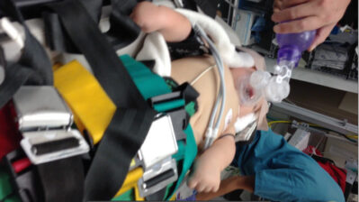
Figure 1 – An infant with Decerebrate Posturing

Exposure
Every trauma patient should be completely exposed early in the resuscitation. Patients should be fully undressed and appropriately log-rolled while maintaining spinal immobilization to look for bruising, lacerations, hemorrhage, entrance/exit wounds, and foreign bodies, when possible. Shortly thereafter, the patient should be covered with warm blankets to prevent hypothermia. Warmed fluids, warmed oxygen, and a warmed resuscitation room can also aid in maintaining normothermia.
Family
The addition of “F” for “family involvement” is recommended to aid in the acute evaluation and provide support to the child. Multiple studies have shown a benefit to both the family and the patient by being present during pediatric resuscitations, regardless of outcome.10 Though the presence of family is sometimes thought of as distracting to providers, having a dedicated staff member (i.e., social worker, chaplain, nurse) who can explain the process and field questions will help keep the resuscitation running smoothly. Observation of family interaction and response is also important, as the possibility of nonaccidental trauma (i.e., abuse) is always a consideration in pediatric trauma.
Secondary Survey
Once life-threatening injuries have been identified and addressed in the primary survey, a secondary head-to-toe exam should be performed to carefully look for additional injuries.5 More detailed descriptions of the evaluations and management of each anatomic area will be included in subsequent sections of this module.
Head
-
Scalp should be examined for hematoma, step-off, bruising around the eyes (raccoon eyes) or laceration that could indicate an intracranial injury or skull fracture.
-
Infants with open fontanels should have an assessment for fullness that may indicate increased intracranial pressure.
-
Ears should be examined for hemotympanum or bruising behind the ear (Battle sign), which could specifically indicate a basilar skull fracture.
-
The face should be assessed for midface stability, jaw alignment, and facial symmetry.
-
The nasopharynx and oropharynx should be examined for bleeding, laceration, hematoma, and dental injury.
-
Eyes should be assessed for pupillary reaction, symmetry, extra-ocular movements, and proptosis. Abnormalities may indicate nerve or muscle entrapment or retrobulbar hemorrhage. Patients with conjunctival injection or irregular pupils should have fluorescein staining and slit lamp examination to assess for corneal injury.
Neck/Spine
-
Palpation for tenderness and step-off on each vertebra for signs of fracture or dislocation.
-
Any neurologic deficit on examination should prompt advanced imaging for fracture, dislocation, and spinal cord injury.
Chest
-
Inspection and palpation for crepitus, tenderness, flail segments, and work of breathing.
-
Auscultation for crackles or decreased breath sounds, which may indicate pulmonary contusion, pneumothorax, or hemothorax.
Cardiac
-
Inspection for perfusion of all extremities.
-
Palpation of pulses and assessment of skin for capillary refill to assess for perfusion. This should be frequently reassessed throughout a resuscitation as the findings may be dynamic.
-
Auscultation for muffled heart sounds, rubs, or gallops which may indicate a pericardial bleeding or cardiac contusion.
Abdomen
-
Inspection for distension, bruising, abrasions, handlebar or seatbelt marks, or other signs of trauma that may indicate intra-abdominal bleeding.
-
Gentle palpation for tenderness, rebound, or peritonitis, the abdominal exam may change over the course of the resuscitation, especially in blunt trauma, and should be repeated as often as necessary.
-
Rectal exam for sphincter tone and gross blood, which may indicate a spinal cord or rectal injury.
Pelvis
-
Genitalia should be inspected for blood at the urethral meatus or perineal bruising. If these are present, bladder catheterization is relatively contraindicated until a retrograde urethrogram demonstrates a normal urethra.
-
Pelvis should be compressed to assess for stability. If the pelvis is unstable, it should be compressed with a pelvic binder or bedsheet.
Extremities
-
Inspection for laceration, bruising, deformity, color, and spontaneous movement.
-
Palpation of pulses. In the case of an obvious deformity without distal pulses, immediate reduction should be performed to restore perfusion.
-
Palpation for tenderness, which may indicate a fracture or growth plate injury. Patients with negative X-rays can still have growth plate injuries, so thorough palpation is important to identify injury.
Ancillary Studies
A detailed history and thorough physical exam is the cornerstone to a good trauma evaluation. Laboratory studies and imaging can help further elucidate specific injuries for treatment and operative plans.
Laboratory tests
After obtaining IV access, several laboratory tests should be considered for both diagnosis and treatment:
-
Type and screen – Preparation of blood for emergent or operative transfusion; consider in patients requiring > 20mL/kg of fluid resuscitation.
-
Complete blood count – Evaluate for degree of hemorrhage and platelet function. Beware that anemia is often not readily apparent in acute hemorrhage. A patient with rapid, sudden blood loss may initially demonstrate a normal hemoglobin and hematocrit. These must be interpreted within the clinical context and are generally best trended over time.
-
Arterial/venous blood gas/lactate – Elevated lactate levels or the presence of a metabolic acidosis with a base deficit can be signs of poor perfusion and can help guide the resuscitation. pH and pCO2 may help assess the patient’s respiratory status and reserve.
-
Transaminases – Elevated transaminases can be early markers of liver injury, although they can also be present in skeletal muscle injury such as a crush mechanism. AST or ALT levels is an effective screening tool for intra-abdominal injury (IAI) in children following blunt abdominal trauma (BAT). Pediatric patients with a negative FAST and liver transaminases < 100 IU/L have a low risk of IAI and may be observed rather than subjected to the radiation risk of CT.11
-
Lipase – With directed blows to the epigastrum, a traumatic pancreatitis can occur in children.
-
Coagulation studies – With severe hemorrhage, patients can develop a coagulopathy after resuscitation. Initial and serial coagulation tests can reveal an emerging coagulopathy that may require various blood products.
Urinalysis
One of the most sensitive indicators and a useful screening tool for renal injury is the presence of hematuria on urinalysis. A normal urinalysis suggests that there is a very low risk of renal injury. The presence of red blood cells (>/=- 50 RBCs per HPF), on the other hand, may prompt further imaging.12
Ultrasound
Pediatric bedside ultrasound has become a valuable adjunct to the trauma assessment. The Focused Assessment Sonography in Trauma (FAST) rapidly obtains three views of the abdomen (RUQ/“Morrison’s pouch,” LUQ, suprapubic) and chest (subxyphoid) to identify signs of intra-abdominal or intra-pericardial fluid.13 Though not a replacement for CT scanning, it is useful in the child who is unstable and cannot be safely transported to the radiology suite.
Figure 2 – A Positive Right Upper Quadrant FAST Exam (blood is seen as a black stripe)

Radiographs
The standard radiographs are a chest X-ray (portable, AP-view), pelvis X-ray (portable, AP-view), and cross table cervical spine X-ray (portable, lateral view). A chest X-ray can quickly identify pneumothoraces, hemothoraces, rib fractures, and other serious chest problems. In pediatric trauma patients, the presence of chest tenderness, back abrasions, or an abnormal respiratory rate have been shown to be associated with the majority of abnormal chest radiographs.14 Routine pediatric pelvic films have been questioned with a screening tool that combines eight high-risk clinical findings (pelvic tenderness, laceration, ecchymosis, abrasion, GCS < 14, positive urinalysis, abdominal pain/tenderness, femur fracture) and five high-risk mechanisms of injury (unrestrained motor vehicle collision [MVC], MVC with ejection, MVC rollover, auto vs pedestrian, auto vs bicycle) recommended to exclude the need for imaging. Absence of all findings accurately predicted the absence of a fracture.15
A cross-table cervical spine X-ray can identify gross abnormalities of the cervical spine such as fracture or dislocation. Spinal tenderness, torticollis, or focal or global neurologic abnormalities should prompt advanced imaging.
The secondary survey should guide further X-ray evaluation, especially with regard to bony tenderness, obvious deformity, or refusal to move a limb. A skeletal survey should be considered in children < 2 years of age in whom abuse is suspected. In an unstable or multiply-injured patient, some studies may need to be deferred until more serious injuries have been addressed or the patient is transferred to a higher level of care.
CT scans
CT scanning is a highly sensitive and specific imaging modality in the setting of trauma. However, it poses the risk of radiation. These risks and benefits must be considered in the individual patient. There are many decision tools that can help to better risk-stratify patients. These will be discussed in subsequent sections of this module.
CT scans of the head and spine can quickly identify intracranial hemorrhage, cerebral edema, and herniation, as well as spinal fractures, dislocation, or subluxation with great accuracy. However, they fail to adequately image neural tissue and can be falsely negative in the setting of diffuse axonal injury and/or spinal cord injury. MRI is recommended for patients with abnormal neurologic exams and normal CT scans.
CT scan of the abdomen identifies solid organ injury with very high sensitivity and specificity. In addition, hollow organ and vascular injuries are readily identified, although with slightly lower sensitivity. Some injuries, such as duodenal hematomas, may develop over time. As a result, even if the initial CT scan is normal, some children may benefit from a period of observation with serial abdominal exams.
CME Question
5. Which of the following findings on primary and secondary survey likely represents a serious injury, even if advanced imaging is normal?
-
Tenderness over a growth plate
-
Abdominal tenderness
-
Asymmetric neurologic exam
-
Perseveration and agitation
-
All of the above
Start or Resume CME Test
(Opens in a new Tab/Window)
References
1. Centers for Disease Control and Prevention. Injury Prevention and Control: Data & Statistics (WISQARS). Available at: www.cdc.gov/injury/wisqars/fatal_injury_reports.html. Accessed December 4, 2014.
2. Romig LE. Pediatric triage. A system to JumpSTART your triage of young patients at MCIs. JEMS 2002;27:52-58. http://www.ncbi.nlm.nih.gov/pubmed/?term=12141119
3. American Academy of Pediatrics and American College of Emergency Physicians. Trauma. In: APLS: The Pediatric Emergency Medicine Resource. 5th edition. Burlington, MA: Jones and Barlett Learning; 2011.
4. Dieckmann RA, Brownstein D, Gausche-Hill M. The pediatric assessment triangle: A novel approach for the rapid evaluation of children. Pediatr Emerg Care 2010;26:312-315. http://www.ncbi.nlm.nih.gov/pubmed/20386420
5. American College of Surgeons. Extremes of Age. A. Pediatric Trauma. In: Advanced Trauma Life Support for Doctors. 7th ed. Chicago: American College of Surgeons; 2004.
6. Kenefake ME, Swarm M, Walthall J. Nuances in pediatric trauma. Emerg Med Clin North Am 2013;31:627-652. http://www.ncbi.nlm.nih.gov/pubmed/23915597
7. King BR, Baker MD, Braitman LE, et al. Endotracheal tube selection in children: A comparison of four methods. Ann Emerg Med 1993;22:530-534.
8. Kleinman ME, Chameides L, Schexnayder SM, et al. Pediatric advanced life support: 2010 American Heart Association guidelines for cardiopulmonary resuscitation and emergency cardiovascular care. Pediatrics 2010;126:e1361-e1399. http://www.ncbi.nlm.nih.gov/pubmed/20956434
9. Simpson D, Reilly P. Pediatric coma scale. Lancet 321982;2:450. http://www.ncbi.nlm.nih.gov/pubmed/6124846
10. Tinsley C, Hill JB, Shah J, et al. Experience of families during cardiopulmonary resuscitation in a pediatric intensive care unit. Pediatrics 2008;122: e799-e804. http://www.ncbi.nlm.nih.gov/pubmed/18829777
11. Sola JE, Cheung MC, Yang R, et al. Pediatric FAST and elevated liver transaminases: An effective screening tool in blunt abdominal trauma. J Surg Res 2009;157:103-107. http://www.ncbi.nlm.nih.gov/pubmed/19592033 http://www.journalofsurgicalresearch.com/article/S0022-4804(09)00145-0/abstract
12. Santucci RA, Langenburg SE, Zachareas MJ. Traumatic hematuria in children can be evaluated as in adults. J Urol 2004;171(2 Pt 1):822-825. http://www.ncbi.nlm.nih.gov/pubmed/14713834
13. Stafford PW, Blinman TA, Nance ML. Practical points in evaluation and resuscitation of the injured child. Surg Clin North Am 2002;82:273-301. http://www.ncbi.nlm.nih.gov/pubmed/12113366
14. Gittelman MA, Gonzalez-del-Rey J, Brody AS, DiGiulio GA. Clinical predictors for the selective use of chest radiographs in pediatric blunt trauma evaluations. J Trauma 2003;55:670-676. http://www.ncbi.nlm.nih.gov/pubmed/14566121
15. Lagisetty J, Slovis T, Thomas R, et al. Are routine pelvic radiographs in major pediatric blunt trauma necessary? Pediatr Radiol 2012;42:853-858. http://www.ncbi.nlm.nih.gov/pubmed/22246414 http://www.researchgate.net/publication/221748710