What one ED physician missed on an ultrasound
What one ED physician missed on an ultrasound
By Staci Kusterbeck, Contributing Editor
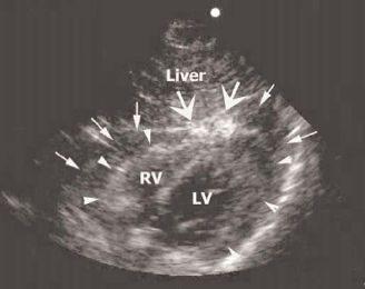
The following ultrasound image of a patient who had been shot in the chest and was not doing well. The physician thought the patient's lung was collapsed, and he put in a chest tube. He then performed an ultrasound of the heart and saw no pericardial effusion and noted that the heart was beating.
 |
| Image courtesy of Michael Blaivas, MD, RDMS, Medical College of Georgia, Augusta. |
"He [the physician] had never taken an ultrasound course and learned a bit here and there on his own," says Michael Blaivas, MD, RDMS, chief of the section of emergency ultrasound in the department of emergency medicine at Medical College of Georgia in Augusta. "He was not credentialed for ultrasound in any way by the hospital as he had never really been that interested in it."
The patient only got worse, but the heart was not involved. The patient was given fluids and blood through central lines but lost his pressure despite this. The ultrasound image shows a mistake that was potentially fatal to this patient.
The ultrasound image is from the subxiphoid position, part of the FAST examination. The liver is labeled and on top of the image. The left ventricle (LV) is seen and so is the right ventricle (RV). "The two atria are not clear in this shot but were seen well in others," explains Blaivas. The long thin arrows point to what was thought to be the outer border of the heart, the outer pericardial layer. The arrow heads point to the actual outer outline of the heart. What is in between? A pericardial effusion that has clotted and looks bright and was confused with being part of the myocardium.
The large arrows point to a bright spot at the apex of the heart. It is not normally seen there. This is where the bullet actually hit. It did not penetrate the heart, but only caused a bruise that led to bleeding into the pericardial sac.
All of this was confirmed at autopsy. If the scan had been interpreted correctly, it would have led to an immediate thorocotomy and cutting of the pericardial sac, which would have revealed tamponade.
"This is Monday morning quarterbacking but is a valid point," says Blaivas. "Had this physician obtained proper training at a course, practiced these scans enough to be credentialed by the hospital, and kept up his skills as with anything in emergency medicine, he would have known to look more carefully, use additional views and [would have] caught the effusion."
An ultrasound image shows a patient who had been shot in the chest and was not doing well. The physician thought the patient's lung was collapsed, and he put in a chest tube.Subscribe Now for Access
You have reached your article limit for the month. We hope you found our articles both enjoyable and insightful. For information on new subscriptions, product trials, alternative billing arrangements or group and site discounts please call 800-688-2421. We look forward to having you as a long-term member of the Relias Media community.
