Scapholunate Dissociation
By William J. Brady, MD
|
Scapholunate dissociation rapidly is becoming the most common form of carpal instability. Despite the literature supporting the importance of early diagnosis and treatment, this problem is not often recognized on initial presentation. With the growing awareness of the problem of occult scaphoid fracture, scapholunate dissociation seems to have replaced this carpal fracture as the most commonly misdiagnosed cause of the wrist sprain diagnosis.1,2 If recognized initially and treated appropriately, this condition has a very good prognosis in terms of recovery of both motion and function. If treated late, however, chronic disability is inevitable.3 On initial presentation, it is vitally important to realize that these patients do not necessarily have tenderness in the anatomic snuffbox and that their symptoms lessen rapidly. As such, if the diagnosis is not made or suspected in the emergency department (ED), the patient may never make it to the orthopedist until the condition becomes chronic and the outcome much less favorable.
These injuries occur with hyperextension and/or the FOOSH (fall-on-an-outstretched-hand) mechanism.4 Subjectively, the complaints of pain vary from minimal to excruciating. Patients often will complain of weakness, and possibly a click or clunk with gripping activity. Additionally, the patient who presents to the ED with complaints of a growing dorsal wrist mass should be evaluated with radiographs, as a dorsal ganglion cyst may be associated with the chronic form of this condition. The most important portion of the physical examination, as with all wrist injuries, is exact localization of the point of maximal tenderness. These patients will localize their maximum tenderness to the scapholunate junction, which is palpable just distal to Lister’s tubercle on the dorsal aspect of the wrist. They may or may not also have snuffbox or tubercle tenderness. Wrist motion is very variable and may be extremely limited or nearly normal. The amount of swelling also is very variable.
 |
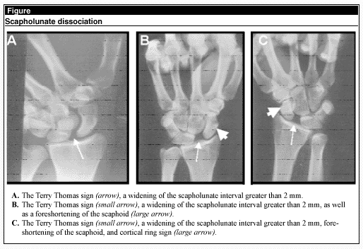
The radiographic findings in this condition are numerous and readily visible on three films. The supinated AP, clenched fist AP, and lateral views will demonstrate the following characteristic findings: 1) widening of the scapholunate gap to greater than 2 mm (Terry Thomas sign) is an indicator of scapholunate dissociation, which is best visualized on a supinated AP or clenched fist AP view, as seen in Figure 1;5 2) foreshortened appearance of the scaphoid in palmar flexed position on the AP view; 3) cortical ring sign seen in the AP view, representing a double density shadow produced by the axial projection of the abnormally oriented scaphoid upon itself; 4) on the lateral view, the scaphoid lies more perpendicular to the axis of the radius rather than at its usual angle of 45-60 degrees; 5) trapezoidal lunate as seen on the AP view is produced by a triangular shape of the lunate as it overlaps the capitate, an indicator of the rotation of the lunate into an extended position; 6) Taleisnik’s "V" sign is produced by palmar flexion of the scaphoid, leading to a more acute angle between the distal radius and scaphoid forming a sharper V-shaped pattern rather than the broad C-shaped arc seen in the normal wrist.
Standard radiographs, however, may be entirely normal. When a scapholunate ligament injury is suspected clinically, additional stress views may be obtained—ulnar deviation with a clenched fist (the clenched fist AP view) will accentuate widening of the scapholunate joint.6
Treatment for this disorder is relatively simple in the ED. A thumb spica splint is applied acutely and referral is made to an orthopedic or hand surgeon for operative repair. This referral is not emergent, though the patient should be seen within 10 days to facilitate early repair. The real key to ensuring appropriate treatment for this potentially disabling condition is to have a high index of suspicion and to make the diagnosis acutely so that early repair is possible.
References
1. Larsen CF, et al. Epidemiology of acute wrist trauma. Int J Epidemiol 1993;22:911-916.
2. Meldon SW, et al. Ligamentous injuries of the wrist. J Emerg Med 1995;13:217-225.
3. Watson HK, et al. The natural progression of scaphoid instability. Hand Clinics 1997;13:39-49.
4. Mayfield JK, et al. Carpal dislocations: Pathomechanics and progressive perilunar instability. J Hand Surg 1980;5:226-241.
5. Frankel VH. The Terry Thomas sign. Clin Orthop 1978;135:311-312.
6. Yeager BA, et al. Radiology of trauma to the wrist: Dislocations, fracture dislocations, and instability patterns. Skeletal Radiol 1985;13:120-130.
Dr. Brady, Associate Professor of Emergency Medicine and Internal Medicine, Residency Director and Vice Chair, Emergency Medicine, University of Virginia, Charlottesville, is on the Editorial Board of Emergency Medicine Alert.
Subscribe Now for Access
You have reached your article limit for the month. We hope you found our articles both enjoyable and insightful. For information on new subscriptions, product trials, alternative billing arrangements or group and site discounts please call 800-688-2421. We look forward to having you as a long-term member of the Relias Media community.
